
MABC949
Anti-Glycoprotein 78 Antibody, clone 3F3A
clone 3F3A, from rat
Sinónimos:
E3 ubiquitin-protein ligase AMFR, AMF receptor, Autocrine motility factor receptor, gp78, RING finger protein 45
About This Item
Productos recomendados


origen biológico
rat
Nivel de calidad
forma del anticuerpo
purified immunoglobulin
tipo de anticuerpo
primary antibodies
clon
3F3A, monoclonal
reactividad de especies
mouse, human
técnicas
immunocytochemistry: suitable
immunohistochemistry: suitable
western blot: suitable
isotipo
IgMκ
Nº de acceso NCBI
Nº de acceso UniProt
Condiciones de envío
ambient
modificación del objetivo postraduccional
unmodified
Información sobre el gen
human ... GPR78(27201)
Descripción general
Especificidad
Inmunógeno
Aplicación
Affects Function: A representative lot stimulated the mobility of B16-F1 mouse melanoma cells on colloidal gold-coated surface (Nabi, I.R., et al. (1990). Cancer Res. 50(2):409-414).
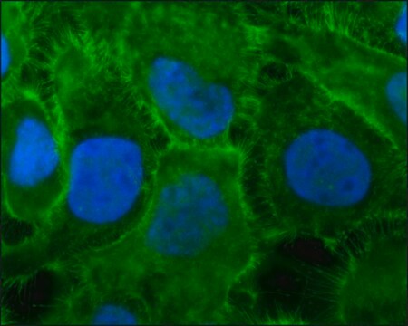
Immunocytochemistry Analysis: A representative lot detected both surface and cytoplasmic gp78 immunoreactivity by fluorescent immunocytochemistry staining of intact or MeOH/3% paraformaldehyde-fixed and 0.5% Triton X-100-permeablized mouse A31 fibroblasts and A31M angiosarcoma cells (Niinaka, Y., et al. (1998). Cancer Res. 58(12):2667-2674).
Immunocytochemistry Analysis: Clone 3F3A ascites fluid immunolocalized gp78 on the surface of adherent B16-F1 mouse melanoma cells by fluorescent immunocytochemistry staining of 3% paraformaldehyde-fixed cells (Nabi, I.R., et al. (1990). Cancer Res. 50(2):409-414).
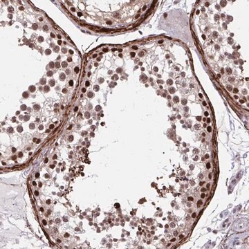
Immunohistochemistry Analysis: A representative lot detected an elevated gp78 immunoreactivity in frozen mammary gland sections from 235RLF transgenic mice than age-matched wild type mice (Joshi, B., et al. (2010). J. Biol. Chem. 285(12):8830-8839).
Western Blotting Analysis: A representative lot detected shRNA-mediated gp78 downregulation in HT-1180 human fibrosarcoma cells (Fu, M., et al. (2013). Mol. Biol. Cell. 24(8):1153-1162).
Western Blotting Analysis: A representative lot detected endogenous gp78 and gp78 transgene expression in mammary glands from wild-type and transgenic mice. Clone 3F3A detected a higher gp78 expression in metastatic MDA-435 cells than in non-metastatic MCF7 breast carcinoma cells (Joshi, B., et al. (2010). J. Biol. Chem. 285(12):8830-8839).
Western Blotting Analysis: A representative lot detected much higher gp78 expression in angiosarcoma (human HT-1180 and mouse A31M) than in fibroblast (human IMR90 and mouse A31) lines (Niinaka, Y., et al. (1998). Cancer Res. 58(12):2667-2674).
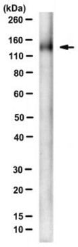
Western Blotting Analysis: A representative lot detected gp78 and a ~150 kDa band in B16-F1 mouse melanoma cell extract. Autocrine motility factor (AMF) competed against clone 3F3A for binding the gp78 target band. Cell extract sialidase treatment decreased the antibody′s immunoreactivity toward gp78 and abolished ~150 kDa band detection (Nabi, I.R., et al. (1990). Cancer Res. 50(2):409-414).
Apoptosis & Cancer
Calidad
Descripción de destino
Forma física
Almacenamiento y estabilidad
Handling Recommendations: Upon receipt and prior to removing the cap, centrifuge the vial and gently mix the solution. Aliquot into microcentrifuge tubes and store at -20°C. Avoid repeated freeze/thaw cycles, which may damage IgG and affect product performance.
Otras notas
Cláusula de descargo de responsabilidad
¿No encuentra el producto adecuado?
Pruebe nuestro Herramienta de selección de productos.
Código de clase de almacenamiento
12 - Non Combustible Liquids
Clase de riesgo para el agua (WGK)
WGK 2
Punto de inflamabilidad (°F)
Not applicable
Punto de inflamabilidad (°C)
Not applicable
Certificados de análisis (COA)
Busque Certificados de análisis (COA) introduciendo el número de lote del producto. Los números de lote se encuentran en la etiqueta del producto después de las palabras «Lot» o «Batch»
¿Ya tiene este producto?
Encuentre la documentación para los productos que ha comprado recientemente en la Biblioteca de documentos.
Nuestro equipo de científicos tiene experiencia en todas las áreas de investigación: Ciencias de la vida, Ciencia de los materiales, Síntesis química, Cromatografía, Analítica y muchas otras.
Póngase en contacto con el Servicio técnico