Saltar al contenido
Merck

SAB4200684

Sigma-Aldrich

Monoclonal Anti-Uvomorulin/E-Cadherin antibody produced in rat

clone DECMA-1, purified from hybridoma cell culture

Sinónimos:

Arc-1, CD324, CDHE, E-cadherin (epithelial), ECAD, LCAM, UVO, cadherin 1, type 1

Iniciar sesiónpara Ver la Fijación de precios por contrato y de la organización


About This Item

UNSPSC Code:
12352209
NACRES:
NA.41

biological source

rat

Quality Level

conjugate

unconjugated

antibody form

purified immunoglobulin

antibody product type

primary antibodies

clone

DECMA-1, monoclonal

form

buffered aqueous solution

mol wt

antigen ~120 kDa

species reactivity

dog, mouse

concentration

~1 mg/mL

technique(s)

immunoblotting: 0.5-1 μg/mL using whole extract of MDCK cells.
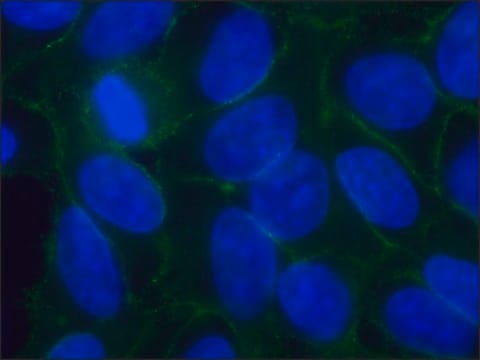
immunofluorescence: 5-10 μg/mL using MDCK cells
immunoprecipitation (IP): suitable

isotype

IgG1

shipped in

dry ice

storage temp.

−20°C

target post-translational modification

unmodified

Gene Information

mouse ... Cdh1(12550)

General description

Monoclonal Anti-Uvomorulin/E-Cadherin (rat IgG1 isotype) is derived from the DECMA-1 hybridoma produced by the fusion of rat myeloma cells and splenocytes from an immunized Lou rat. Uvomorulin/E-Cadherin (Epithelial cadherin), also known as CDH1, CAM 120/80, L-CAM (Cell-Adhesion Molecule) or CD324, is a member of the cadherin transmembrane glycoproteins superfamily. It is characterized with a highly conserved cytoplasmic tail.

Immunogen

Mouse embryonal carcinoma cell line PCC4 Aza RI

Application

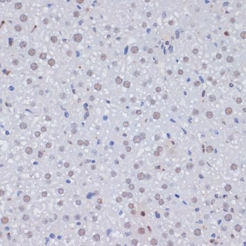
Monoclonal Anti-Uvomorulin/E-Cadherin antibody produced in rat has been used in various immunochemical techniques including immunoblotting, immunoprecipitation, immunohistochemical staining and immunofluorescent staining.

Biochem/physiol Actions

Cadherins contribute to epithelial cell behavior and tissue morphogenesis/remodeling by facilitating cell-cell adhesion. Monoclonal Anti-Uvomorulin/E-Cadherin can be used for studies of embryonal development, cell-cell interaction of cells grown in culture, and localization of E-Cadherin. In addition, it is commonly used as an E-cadherin inhibitor/blocking antibody in a study of embryonic stem cells (EMS).
E-cadherin is a calcium-dependent glycoprotein. It is responsible for the homogeneous attachment between cells. It suppresses malignant transformation of tumors. Additionally, it also regulates cellular invasion and metastasis.

Physical form

Solution in 0.01 M phosphate buffered saline, pH 7.4, containing 15 mM sodium azide.

Disclaimer

Unless otherwise stated in our catalog or other company documentation accompanying the product(s), our products are intended for research use only and are not to be used for any other purpose, which includes but is not limited to, unauthorized commercial uses, in vitro diagnostic uses, ex vivo or in vivo therapeutic uses or any type of consumption or application to humans or animals.

¿No encuentra el producto adecuado?  

Pruebe nuestro Herramienta de selección de productos.

Storage Class

10 - Combustible liquids

flash_point_f

Not applicable

flash_point_c

Not applicable


Certificados de análisis (COA)

Busque Certificados de análisis (COA) introduciendo el número de lote del producto. Los números de lote se encuentran en la etiqueta del producto después de las palabras «Lot» o «Batch»

¿Ya tiene este producto?

Encuentre la documentación para los productos que ha comprado recientemente en la Biblioteca de documentos.

Visite la Librería de documentos

Adherens and tight junctions: structure, function and connections to the actin cytoskeleton
Hartsock A and Nelson WJ
Biochimica et Biophysica Acta - Biomembranes, 1778(3), 660-669 (2008)
Cadherin-mediated cell-cell interactions in normal and cancer cells
Gloushankova NA, et al.
Tissue barriers, 5(3), e1356900-e1356900 (2017)
Kento Takaya et al.
Scientific reports, 14(1), 16442-16442 (2024-07-17)
Wounds that occur in adults form scars due to fibrosis, whereas those in embryos regenerate. If wound healing in embryos is mimicked in adults, scarring can be reduced. We found that mouse fetuses could regenerate tissues up to embryonic day
Kento Takaya et al.
Scientific reports, 12(1), 15913-15913 (2022-09-24)
Up to a certain developmental stage, a fetus can completely regenerate wounds in the skin. To clarify the mechanism of fetal skin regeneration, identifying when the skin switches from fetal-type wound regeneration to adult-type wound repair is necessary. We hypothesized
Yan Zhang et al.
Molecular medicine reports, 12(3), 3393-3399 (2015-05-16)
The aim of the present study was to investigate the expression and clinical significance of hypoxia-inducible factor 1α (HIF-1α), Snail and E-cadherin in ovarian cancer. The expression levels were assessed in a number of ovarian cancer cell lines and ovarian

Nuestro equipo de científicos tiene experiencia en todas las áreas de investigación: Ciencias de la vida, Ciencia de los materiales, Síntesis química, Cromatografía, Analítica y muchas otras.

Póngase en contacto con el Servicio técnico