Saltar al contenido
Merck
Todas las fotos(1)

Documentos clave

369A-1

Sigma-Aldrich

Phosphohistone H3 (PHH3) Rabbit Polyclonal Antibody

Iniciar sesiónpara Ver la Fijación de precios por contrato y de la organización


About This Item

Código UNSPSC:
12352203
NACRES:
NA.41

origen biológico

rabbit

Nivel de calidad

100
500

conjugado

unconjugated

forma del anticuerpo

Ig fraction of antiserum

tipo de anticuerpo

primary antibodies

clon

polyclonal

descripción

For In Vitro Diagnostic Use in Select Regions (See Chart)

Formulario

buffered aqueous solution

reactividad de especies

human

envase

vial of 0.1 mL concentrate (369A-14)
vial of 0.5 mL concentrate (369A-15)
bottle of 1.0 mL predilute (369A-17)
vial of 1.0 mL concentrate (369A-16)
bottle of 7.0 mL predilute (369A-18)

fabricante / nombre comercial

Cell Marque®

técnicas

immunohistochemistry (formalin-fixed, paraffin-embedded sections): 1:100-1:500
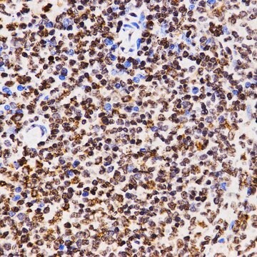
control

tonsil

Condiciones de envío

wet ice

temp. de almacenamiento

2-8°C

visualización

nuclear

Información sobre el gen

human ... H3C1(8350)

Descripción general

Phosphohistone H3 (PHH3) is a core histone protein, which together with other histones, forms the major protein constituents of the chromatin in eukaryotic cells. In mammalian cells, phosphohistone H3 is negligible during interphase but reaches a maximum for chromatin condensation during mitosis. Immunohistochemical studies showed anti-PHH3 specifically detected the core protein histone H3 only when phosphorylated at serine 10 or serine 28. Studies have also revealed no phosphorylation on the histone H3 during apoptosis. Therefore, PHH3 can serve as a mitotic marker to separate mitotic figures from apoptotic bodies and karyorrhectic debris, which may be a very useful tool in the diagnosis of tumor grades, especially in CNS, skin, gyn., soft tissue, and GIST.

Calidad


IVD

IVD

IVD

RUO

Ligadura / enlace

Phosphohistone H3 (PHH3) Positive Control Slides, Product No. 369S, are available for immunohistochemistry (formalin-fixed, paraffin-embedded sections).

Forma física

Solution in Tris Buffer, pH 7.3-7.7, with 1% BSA and <0.1% Sodium Azide

Nota de preparación

Download the IFU specific to your product lot and formatNote: This requires a keycode which can be found on your packaging or product label.

Otras notas

For Technical Service please contact: 800-665-7284 or email: [email protected]

Información legal

Cell Marque is a registered trademark of Merck KGaA, Darmstadt, Germany

¿No encuentra el producto adecuado?  

Pruebe nuestro Herramienta de selección de productos.


Elija entre una de las versiones más recientes:

Certificados de análisis (COA)

Lot/Batch Number

¿No ve la versión correcta?

Si necesita una versión concreta, puede buscar un certificado específico por el número de lote.

¿Ya tiene este producto?

Encuentre la documentación para los productos que ha comprado recientemente en la Biblioteca de documentos.

Visite la Librería de documentos

Sayantani Goswami et al.
Journal of cellular physiology, 236(12), 8148-8159 (2021-07-01)
A number of studies have examined the effects of 1,25-dihydroxyvitamin D3 (1,25(OH)2 D3 ) on intestinal inflammation driven by immune cells, while little information is currently available about its impact on inflammation caused by intestinal epithelial cell (IEC) defects. Mice
M J Hendzel et al.
The Journal of biological chemistry, 273(38), 24470-24478 (1998-09-12)
Apoptosis plays an important role in the survival of an organism, and substantial work has been done to understand the signaling pathways that regulate this process. Characteristic changes in chromatin organization accompany apoptosis and are routinely used as markers for
Histone phosphorylation and chromatin structure during mitosis in Chinese hamster cells.
L R Gurley et al.
European journal of biochemistry, 84(1), 1-15 (1978-03-01)
Howard Colman et al.
The American journal of surgical pathology, 30(5), 657-664 (2006-05-16)
Distinguishing between grade II and grade III diffuse astrocytomas is important both for prognosis and for treatment decision-making. However, current methods for distinguishing between grades based on proliferative potential are suboptimal, making identification of clear cutoffs difficult. In this study
Yoo-Jin Kim et al.
American journal of clinical pathology, 128(1), 118-125 (2007-06-21)
Mitotic activity is one of the most reliable prognostic factors in meningiomas. The identification of mitotic figures (MFs) and the areas of highest mitotic activity in H&E-stained slides is a tedious and subjective task. Therefore, we compared the results from

Nuestro equipo de científicos tiene experiencia en todas las áreas de investigación: Ciencias de la vida, Ciencia de los materiales, Síntesis química, Cromatografía, Analítica y muchas otras.

Póngase en contacto con el Servicio técnico