Saltar al contenido
Merck
Todas las fotos(1)

Key Documents

168M-9

Sigma-Aldrich

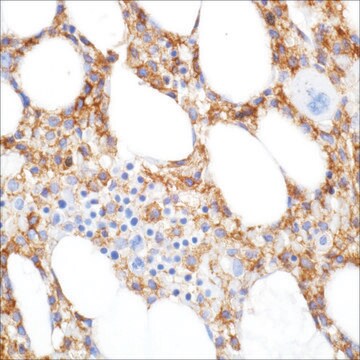
CD68 (Kp-1) Mouse Monoclonal Antibody

Iniciar sesiónpara Ver la Fijación de precios por contrato y de la organización


About This Item

UNSPSC Code:
12352203
NACRES:
NA.41

biological source

mouse

Quality Level

100
500

conjugate

unconjugated

antibody form

culture supernatant

antibody product type

primary antibodies

clone

Kp-1, monoclonal

description

For In Vitro Diagnostic Use in Select Regions (See Chart)

form

buffered aqueous solution

species reactivity

human

packaging

vial of 0.1 mL concentrate (168M-94)
vial of 0.5 mL concentrate (168M-95)
bottle of 1.0 mL predilute (168M-97)
vial of 1.0 mL concentrate (168M-96)
bottle of 7.0 mL predilute (168M-98)

manufacturer/tradename

Cell Marque

technique(s)

immunohistochemistry (formalin-fixed, paraffin-embedded sections): 1:100-1:500

isotype

IgG1κ

control

tonsil

shipped in

wet ice

storage temp.

2-8°C

visualization

cytoplasmic

Gene Information

human ... CD68(968)

General description

Anti-CD68 marks cells of monocyte/macrophage lineage. This antibody is capable of staining monocytes, Kupffer cells, osteoclasts, granulocytes and their precursors; lymphomas are negative or show a few granules. This antibody may be useful for the identification of myelomonocytic and histiocytic tumors. Anti-CD68 may help to distinguish malignant fibrous histiocytoma from other pleomorphic sarcomas. However, since this detects a formalin-resistant epitope that may be associated with lysosomal granules, other lysosome-rich cells may also stain.

Quality


IVD

IVD

IVD

RUO

Linkage

CD68 Positive Control Slides, Product No. 168S, are available for immunohistochemistry (formalin-fixed, paraffin-embedded sections).

Physical form

Solution in Tris Buffer, pH 7.3-7.7, with 1% BSA and <0.1% Sodium Azide

Preparation Note

Download the IFU specific to your product lot and formatNote: This requires a keycode which can be found on your packaging or product label.

Other Notes

For Technical Service please contact: 800-665-7284 or email: service@cellmarque.com

Legal Information

Cell Marque is a trademark of Merck KGaA, Darmstadt, Germany

¿No encuentra el producto adecuado?  

Pruebe nuestro Herramienta de selección de productos.


Certificados de análisis (COA)

Busque Certificados de análisis (COA) introduciendo el número de lote del producto. Los números de lote se encuentran en la etiqueta del producto después de las palabras «Lot» o «Batch»

¿Ya tiene este producto?

Encuentre la documentación para los productos que ha comprado recientemente en la Biblioteca de documentos.

Visite la Librería de documentos

J L Snow et al.
The British journal of dermatology, 133(1), 71-76 (1995-07-01)
The clinical, histopathological, and immunophenotypic characteristics of four cases of malignancy-associated multicentric reticulohistiocytosis (MMR) and one case each of diffuse cutaneous reticulohistiocytosis (DCR) and isolated reticulohistiocytoma (IR), are reviewed. In all four cases of MMR the cutaneous lesions and joint
F Facchetti et al.
Histopathology, 19(2), 141-145 (1991-08-01)
The monoclonal antibody KP1, which recognizes the CD 68 antigen on macrophages and myeloid precursors, was tested on 28 malignant (primary and metastatic) melanomas, 28 naevi, and 17 skin biopsies showing either normal (10) or hyperplastic melanocytes (7). Sixteen of
L P Ruco et al.
American journal of clinical pathology, 92(3), 273-279 (1989-09-01)
The expression of macrophage antigens KP1, Mac, lysozyme, and alpha-1-antichymotrypsin was investigated on routine paraffin sections from 17 cases of Langerhans' cell histiocytosis (LCH). All the major clinical forms were represented, including single lesions and monosystemic and multisystemic disease. In
CD68 cluster workshop report
JL Cordell, et al.
Leucocyte Typing V, 925-927 (1995)
B Vergier et al.
Blood, 95(7), 2212-2218 (2000-03-25)
The course of mycosis fungoides (MF) is indolent except when transformation to a large T-cell lymphoma occurs. The diagnosis of transformed MF (T-MF) relies on the presence of more than 25% of large cells on biopsy of an MF lesion.

Nuestro equipo de científicos tiene experiencia en todas las áreas de investigación: Ciencias de la vida, Ciencia de los materiales, Síntesis química, Cromatografía, Analítica y muchas otras.

Póngase en contacto con el Servicio técnico