Saltar al contenido
Merck
Todas las fotos(1)

Key Documents

MAB4310X

Sigma-Aldrich

Anti-CD133 Antibody, clone 13A4, Alexa Fluor 488 conjugated

clone 13A4, Chemicon®, from rat

Sinónimos:

Prominin-1, AC133

Iniciar sesiónpara Ver la Fijación de precios por contrato y de la organización


About This Item

UNSPSC Code:
12352203
eCl@ss:
32160702
NACRES:
NA.44

biological source

rat

Quality Level

conjugate

ALEXA FLUOR 488

antibody form

purified antibody

antibody product type

primary antibodies

clone

13A4, monoclonal

species reactivity

mouse

should not react with

Drosophila, rat, chicken, human

manufacturer/tradename

Chemicon®

technique(s)

flow cytometry: suitable

input

sample type epithelial cells
sample type hematopoietic stem cell(s)
sample type: mouse embryonic stem cell(s)
sample type mesenchymal stem cell(s)
sample type neural stem cell(s)

isotype

IgG1κ

NCBI accession no.

UniProt accession no.

shipped in

wet ice

target post-translational modification

unmodified

Gene Information

chicken ... Prom1(422825)
human ... PROM1(8842)
mouse ... Prom1(19126)
rat ... Prom1(60357)

General description

Prominin is a novel plasma membrane protein with an N-terminal extracellular domain, five transmembrane segments flanking two short cytoplasmic loops and two large glycosylated extracellular domains, and a cytoplasmic C-terminal domain. DNA sequences from Caenorhabditis elegans predict the existence of a protein with the same features, suggesting that prominin is conserved between vertebrates and invertebrates. Prominin is found not only in the neuroepithelium but also in various other epithelia of the mouse embryo. In the adult mouse, prominin has been detected in the brain ependymal layer, and in kidney tubules. In these epithelia, prominin is specific to the apical surface, where it is selectively associated with microvilli and microvilli-related structures. Remarkably, upon expression in CHO cells, prominin is preferentially localized to plasma membrane protrusions such as filopodia, lamellipodia, and microspikes. These observations imply that prominin contains information to be targeted to, and/or retained in, plasma membrane protrusions rather than the planar cell surface. Moreover, results show that the mechanisms underlying targeting of membrane proteins to microvilli of epithelial cells and to plasma membrane protrusions of non-epithelial cells are highly related (Weigmann et al., 1997). Also see (http://www.ncbi.nlm.nih.gov/prow/guide/438375806_g.htm) for more information on CD133 and prominin expression.

Specificity

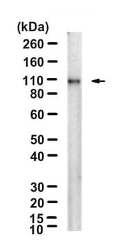
Recognizes mouse Prominin-1, a pentaspan transmembrane (5-TM) domain glycoprotein. Immunoblotting of E12 mouse brain and adult mouse kidney using mAb 13A4 showed that prominin has a mean apparent molecular weight of 115 kDa; Deglycosylation of brain and kidney prominin with peptide N-glycosidase F (PNGase F) yielded a 94-kDa band indicating that prominin is N-glycosylated. The additional 88-kDa band seen upon deglycosylation of brain prominin represents a C-terminally truncated form (Weigmann et al., 1997; Maw et al., 2000; Roper I, 2000). Prominin-1 is expressed on primitive cells such as hematopoietic stem and progenitor cells, neural & endothelial stem cells, retina and retinoblastoma, as well as developing epithelium. To date, the function and ligand of Prominin-1 are unknown. This antibody can be used to isolate murine stem cells from brain and bone marrow.

Immunogen

Telencephali of ten 12-day-old NMRI mouse embryos (E12, ~1 mg protein), which consist mostly of neuroepithelium, were homogenized in 1 mL PBS, mixed with crushed nitrocellulose filter (1 cm2 in 0.5 mL PBS) as adjuvant, and injected i.p. into a LouXSD rat (Weigmann et al., PNAS 1997).

Application

Immunohistochemistry: 1-15 μg/mL. Suggested fixatives: 2-4% PFA, or methanol -20°C fixation. When fixing cells in culture, incubate sample in 3% PFA for 15-30 min at room temperature. Tissue: perfusion with 2-4% PFA, 1-4 hours postfixation is typical. Traditional formalin fixation is NOT recommended. Permeabilization method: 0.2% saponin or 0.1-0.3% Triton X-100 in PBS. Blocking Buffer: For cells in culture, 0.2% gelatin in PBS; for tissue section, 10% FCS in PBS. Dilution Buffer: For cells in culture, 0.2% gelatin in PBS; for tissue section, 0.2% saponin and 10% FCS in PBS. Incubation Times/Temperature: Overnight at 4°C or 1 hour at 37°C.
Note: This antibody does not work with paraffin-embedded sections.

EM immunohistochemistry: The subcellular localization of the 13A4 antigen in mouse E9-10 neuroepithelial cells and adult kidney proximal tubule cells was investigated by immunogold electron microscopy. Strong labeling was observed over the kidney brush border membrane, where 13A4 immunoreactivity appeared to be concentrated toward the tips of the microvilli. Remarkably, in neuroepithelial cells, whose apical plasma membrane contains fewer microvilli than the kidney brush border, 13A4 immunoreactivity was associated mostly, if not exclusively, with microvilli and plasma membrane protrusions, and was not detected in the planar areas of the apical plasma membrane. Because of this preferential localization, the 13A4 antigen was referred to as ′′prominin′′ (from the Latin word ′′prominere,′′ to stand out, to be prominent) (Weigmann et al., 1997).

Western blotting: 1-5 μg/mL in 0.3% Tween in PBS. Sample preparation: Standard Laemmli (boiled in 2% SDS, 100mM DTT or 5% beta-mercaptoethanol, 60mM Tris-HCL pH 6.8). Preferred Gel percentage: 7.5%. Suggested Blocking Buffer: 3-5% Milk, 0.3% Tween in PBS. Incubation time: 1 hour at room temperature or overnight at 4°C. Recommended control extracts: Positive: Kidney membrane; Negative: liver membranes.

Immunoprecipitation: 10-25 μg/mL. Suggested tissue/cell lysis buffer: RIPA bufferFinal reaction volume: 500-1000 μL. Final total protein concentration in reaction mix: 0.5-3 mg/mL. Incubation times: overnight at 4°C. Capture agent used: Protein G Sepharose or rabbit anti-rat antibody/protein A Sepharose. Expected sizes on immunoblots (in kDa): 115 kDa (mature form) or 105 kDa (precursor form).

FACS Analysis: Suggested dilution/number of cells: 0.25-1 μg/ million cells. Fixation/Permeabilization used: BD FACS lysis Solution (1-1.5% formaldehyde) (BD and FACS are trademarks of Becton, Dickinson and Company) No permeabilization. Recommended controls: Hematopoietic stem cells.

Optimal working dilutions must be determined by the end user.
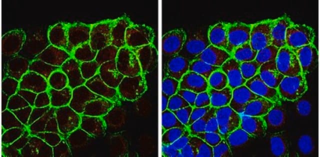
Anti-CD133 Antibody, clone 13A4, Alexa Fluor 488 conjugated is an antibody against CD133 for use in FC.
Research Category
Stem Cell Research
Research Sub Category
Neural Stem Cells

Hematopoietic Stem Cells

Physical form

Liquid in Phosphate buffer with 15 mg/mL BSA as a stabilizer and 0.1% sodium azide.
Protein A purified

Storage and Stability

Maintain refrigerated at 2–8°C protected from light in undiluted aliquots for up to 24 months.

Analysis Note

Control
Human embryonic stem cell lysate, Caco 2 (Human colonic carcinoma cell line) whole cell lysate

Other Notes

Concentration: Please refer to the Certificate of Analysis for the lot-specific concentration.

Legal Information

ALEXA FLUOR is a trademark of Life Technologies
CHEMICON is a registered trademark of Merck KGaA, Darmstadt, Germany
Sepharose is a trademark of Cytiva

Disclaimer

Alexa Fluor is a registered trademark of Molecular Probes, Inc.

Unless otherwise stated in our catalog or other company documentation accompanying the product(s), our products are intended for research use only and are not to be used for any other purpose, which includes but is not limited to, unauthorized commercial uses, in vitro diagnostic uses, ex vivo or in vivo therapeutic uses or any type of consumption or application to humans or animals.

¿No encuentra el producto adecuado?  

Pruebe nuestro Herramienta de selección de productos.

Storage Class

12 - Non Combustible Liquids

wgk_germany

WGK 2

flash_point_f

Not applicable

flash_point_c

Not applicable


Certificados de análisis (COA)

Busque Certificados de análisis (COA) introduciendo el número de lote del producto. Los números de lote se encuentran en la etiqueta del producto después de las palabras «Lot» o «Batch»

¿Ya tiene este producto?

Encuentre la documentación para los productos que ha comprado recientemente en la Biblioteca de documentos.

Visite la Librería de documentos

Margarita Pustovalova et al.
International journal of molecular sciences, 23(9) (2022-05-15)
Cancer stem cells (CSCs) play a critical role in the initiation, progression and therapy relapse of many cancers including non-small cell lung cancer (NSCLC). Here, we aimed to address the question of whether the FACS-sorted CSC-like (CD44 + &CD133 +)

Nuestro equipo de científicos tiene experiencia en todas las áreas de investigación: Ciencias de la vida, Ciencia de los materiales, Síntesis química, Cromatografía, Analítica y muchas otras.

Póngase en contacto con el Servicio técnico