Saltar al contenido
Merck
Todas las fotos(2)

Key Documents

ABC102

Sigma-Aldrich

Anti-CCN1/CYR61 Antibody

from rabbit, purified by affinity chromatography

Sinónimos:

Protein CYR61, CCN family member 1, Cysteine-rich angiogenic inducer 61, Insulin-like growth factor-binding protein 10, IBP-10, IGF-binding protein 10, IGFBP-10, Protein GIG1

Iniciar sesiónpara Ver la Fijación de precios por contrato y de la organización


About This Item

UNSPSC Code:
12352203
eCl@ss:
32160702
NACRES:
NA.41

biological source

rabbit

Quality Level

antibody form

affinity isolated antibody

antibody product type

primary antibodies

clone

polyclonal

purified by

affinity chromatography

species reactivity

human, mouse, rat

species reactivity (predicted by homology)

chimpanzee (based on 100% sequence homology)

technique(s)

immunohistochemistry: suitable (paraffin)
western blot: suitable

NCBI accession no.

UniProt accession no.

shipped in

wet ice

target post-translational modification

unmodified

Gene Information

human ... CYR61(3491)

General description

CCN1 (cysteine-rich 61 or CYR61), a ubiquitous angiogenic factor, is a member of the CNN family, which is also comprised of CTGF, WISP-1, WISP-2, and WISP-3. It is a multidomain protein, containing one each of CTCK, IGFBP, TSP type-1, and VWFC domains. CCN1 is associated with the extracellular matrix and functions as a ligand for integrin receptors. It is expressed in response to growth factor stimulation, and mediates various cellular processes, including cell adhesion, migration, proliferation, angiogenesis, extracellular-matrix remodeling, and placental development. Previous studies have indicated that it may also play a role in wound healing by influencing the expression of genes required for this process, such as TIMP1, PAI-1, VEGA-A, VEGA-C, MMP1, MMP3, and uPA. CCN1 has also been implicated in breast cancer.

Specificity

This antibody recognizes the CTCK domain of: CCN1/CYR61

Immunogen

Epitope: CTCK domain
KLH-conjugated linear peptide corresponding to the CTCK domain of human CCN1/CYR61.

Application

Detect CCN1/CYR61 using this Anti-CCN1/CYR61 Antibody validated for use in Western Blotting, IHC(P).
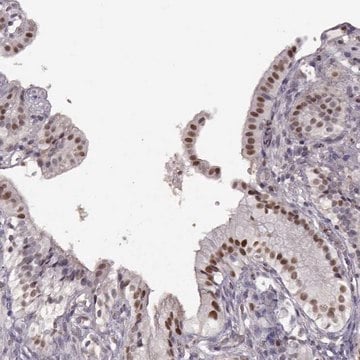
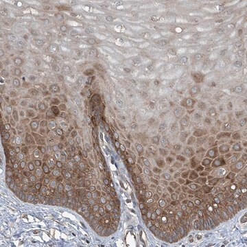
Immunohistochemistry Analysis: 1:200 dilution from a representative lot detected CCN1/CYR61 in human kidney tissue.
Research Category
Apoptosis & Cancer
Research Sub Category
Apoptosis - Additional

Quality

Evaluated by Western Blot in NIH/3T3 cell lysate.

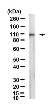
Western Blot Analysis: 2 µg/mL of this antibody detected CCN1/CYR61 in 10 µg of NIH/3T3 cell lysate.

Target description

~53 kDa observed. The calculated molecular weight of this protein is 42 kDa, but can be observed at higher molecular weights due to glycosylation. A degradation fragment may be observed at ~15 kDa in some lysates. (Rother et al., 2010)

Physical form

Antigen Affinity Purified
Purified rabbit polyclonal in buffer containing 0.1 M Tris-Glycine (pH 7.4), 150 mM NaCl with 0.05% sodium azide.

Storage and Stability

Stable for 1 year at 2-8°C from date of receipt.

Analysis Note

Control
NIH/3T3 cell lysate

Other Notes

Concentration: Please refer to the Certificate of Analysis for the lot-specific concentration.

Disclaimer

Unless otherwise stated in our catalog or other company documentation accompanying the product(s), our products are intended for research use only and are not to be used for any other purpose, which includes but is not limited to, unauthorized commercial uses, in vitro diagnostic uses, ex vivo or in vivo therapeutic uses or any type of consumption or application to humans or animals.

¿No encuentra el producto adecuado?  

Pruebe nuestro Herramienta de selección de productos.

Storage Class

12 - Non Combustible Liquids

wgk_germany

WGK 1

flash_point_f

Not applicable

flash_point_c

Not applicable


Certificados de análisis (COA)

Busque Certificados de análisis (COA) introduciendo el número de lote del producto. Los números de lote se encuentran en la etiqueta del producto después de las palabras «Lot» o «Batch»

¿Ya tiene este producto?

Encuentre la documentación para los productos que ha comprado recientemente en la Biblioteca de documentos.

Visite la Librería de documentos

James Hutchenreuther et al.
Cancer research communications, 4(2), 556-570 (2024-02-16)
Melanoma is the leading cause of skin cancer-related death. As prognosis of patients with melanoma remains problematic, identification of new therapeutic targets remains essential. Matricellular proteins are nonstructural extracellular matrix proteins. They are secreted into the tumor microenvironment to coordinate
Weihua Li et al.
Molecular medicine reports, 12(3), 3359-3364 (2015-06-06)
MicroRNAs (miRNAs) are known to regulate the expression of a variety of genes, which are important in the development of several types of tumor, including Ewing's sarcoma (ES), at the post‑transcriptional level. Although previous studies have identified that the expression
Metformin improves tendon degeneration by blocking translocation of HMGB1 and suppressing tendon inflammation and senescence in aging mice.
Zhang, et al.
Journal of Orthopaedic Research : Official Publication of the Orthopaedic Research Society, 41, 1162-1176 (2023)
Tanguy Lechertier et al.
Nature communications, 11(1), 2810-2810 (2020-06-06)
The overexpression of the protein tyrosine kinase, Focal adhesion kinase (FAK), in endothelial cells has implicated its requirement in angiogenesis and tumour growth, but how pericyte FAK regulates tumour angiogenesis is unknown. We show that pericyte FAK regulates tumour growth

Nuestro equipo de científicos tiene experiencia en todas las áreas de investigación: Ciencias de la vida, Ciencia de los materiales, Síntesis química, Cromatografía, Analítica y muchas otras.

Póngase en contacto con el Servicio técnico