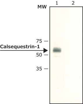
synthetic peptide corresponding to amino acids 28-45 located near the N-terminus of mouse calsequestrin-1, conjugated to KLH. This sequence is identical in rat and highly conserved in human and bovine calsequestrin-1 (two amino acid substitution). This sequence is not found in calsequestrin-2.
Application
Anti-Calsequestrin-1 (N-terminal) antibody produced in rabbit is suitable for western blotting at a concentration of 0.5-1μg/mL using extracts of rat skeletal muscle S1 fraction.
Biochem/physiol Actions
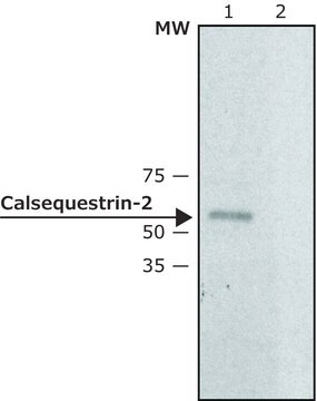
Calsequestrin (CS) is a calcium-binding protein in the sarcoplasmic reticulum (SR). It is known as CSQ and PDIB1. The gene belongs to calsequestrin protein family. Calsequestrin-1 is a putative auto-antigen associated with eye muscle inflammation in Graves′ disease. Calsequestrin-2 (55 kDa) is found in the SR′s terminal cisternae luminal space of cardiac, slow skeletal muscle cells and may be in platelets. The major Ca2+ binding protein in cardiac and skeletal muscle is a high-capacity, low-affinity Ca2+ binding glycoprotein, which functions as an internal Ca2+ store in the lumen of the SR. It helps in controlling the smooth-muscle contraction.
Target description
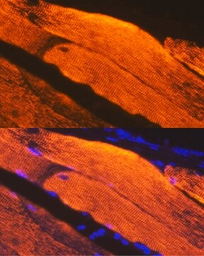
Calsequestrin (CS also known as CSQ), the major Ca2+ binding protein in cardiac and skeletal muscle, is a high-capacity, low-affinity Ca2+ binding glycoprotein, which functions as an internal Ca2+ store in the lumen of the SR. In mammalians, two forms of the protein are found, calsequestrin-1 (CASQ-1) and calsequestrin-2 (CASQ-2), which encode the fast-twitch skeletal muscle and cardiac calsequestrin, respectively. Calsequestrin-1 is found in the SRs terminal cisternae luminal space of fast skeletal muscle cells. Calsequestrin-1 is a putative autoantigen associated with eye muscle inflammation in Graves′ disease.
Physical form
Solution in 0.01 M phosphate buffered saline, pH 7.4, containing 15 mM sodium azide.
Disclaimer
Unless otherwise stated in our catalog or other company documentation accompanying the product(s), our products are intended for research use only and are not to be used for any other purpose, which includes but is not limited to, unauthorized commercial uses, in vitro diagnostic uses, ex vivo or in vivo therapeutic uses or any type of consumption or application to humans or animals.
A 63 kDa skeletal muscle protein associated with eye muscle inflammation in Graves' disease is identified as the calcium binding protein calsequestrin.
We have investigated expression of skeletal calsequestrin (CSQ1) and fiber type composition in normal and regenerated fast and slow skeletal muscles and in the left heart ventricles of euthyroid (EU), hypothyroid (HY) and hyperthyroid (TH) adult inbred Lewis strain rats.
The Journal of cell biology, 98(4), 1597-1602 (1984-04-01)
Localization of calsequestrin in chicken ventricular muscle cells was determined by indirect immunofluorescence and immuno-Protein A-colloidal gold labeling of cryostat and ultracryotomy sections, respectively. Calsequestrin was localized in the lumen of peripheral junctional sarcoplasmic reticulum, as well as in the
Clinical and experimental immunology, 145(1), 56-62 (2006-06-24)
We have identified several eye muscle antigens and studied the significance of the corresponding serum autoantibodies in patients with Graves' disease. Of these antigens, only calsequestrin is expressed more in eye muscle than other skeletal muscles, which could explain at
Journal of thrombosis and haemostasis : JTH, 10(1), 116-124 (2011-11-09)
Altered Ca(2+) homeostasis contributes significantly to platelet hyperactivity in diabetes mellitus. Calsequestrin (CSQ), as a Ca(2+) buffer protein in the sarcoplasmic reticulum, also regulates the Ca(2+) release process in muscles. We hypothesized that CSQ may be expressed in platelets, but
Our team of scientists has experience in all areas of research including Life Science, Material Science, Chemical Synthesis, Chromatography, Analytical and many others.