Skip to Content
Merck
All Photos(1)

Key Documents

C9972

Sigma-Aldrich

Cholera Toxin B subunit

biotin conjugate, lyophilized powder

Synonym(s):

CTB

Sign Into View Organizational & Contract Pricing


About This Item

MDL number:
UNSPSC Code:
12352200
PubChem Substance ID:
NACRES:
NA.77

conjugate

biotin conjugate

Quality Level

form

lyophilized powder

mol wt

~12 kDa

composition

Protein, ~40% Lowry

storage temp.

2-8°C

SMILES string

CCOc1ccccc1C(=O)Nc2ccc(Cl)c(c2)C(F)(F)F

InChI

1S/C16H13ClF3NO2/c1-2-23-14-6-4-3-5-11(14)15(22)21-10-7-8-13(17)12(9-10)16(18,19)20/h3-9H,2H2,1H3,(H,21,22)

InChI key

YDXZSNHARVUYNM-UHFFFAOYSA-N

Looking for similar products? Visit Product Comparison Guide

General description

Cholera Toxin B (CTB) is secreted by Vibrio cholerae. CTB functions as an oral subunit vaccine for cholera, which is associated with acute watery diarrhoea. It acts as a mucosal immunogen. Cholera toxin (CT) stimulates cell surface molecules, such as antigen presenting cells (APCs), murine and human dendritic cells (DCs). CT also has immunomodulatory properties. It induces the secretion of interleukin 1 (IL-1) from macrophages and enhances their APC function.

Application

Cholera Toxin B subunit has been used:
  • in immunofluorescence
  • in the analysis of major histocompatibility complex (MHC) class II lipid raft partitioning
  • in live cell three-dimensional tracking of SH-SY5Y human neuroblastoma cells
  • to assess the toll-like receptors (TLR) and FcRγ (Fc receptor γ chain) – CARD9 (caspase recruitment domain family member 9) activation by cholera Toxin B (CTB)

Biochem/physiol Actions

The cholera toxin B subunit is used for track tracing in neurological research, taking advantage of GM1 ganglioside binding and retrograde transport. Tissue culture cells treated with cholera toxin are not killed and tissues of animals do not become necrotic.

Quality

Biotin content ~1.0 mole/mole protein.

Physical form

Lyophilized powder containing sodium phosphate buffer salts, sodium azide and sodium EDTA.

Analysis Note

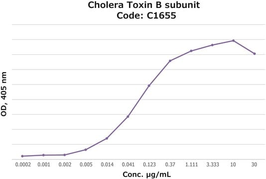
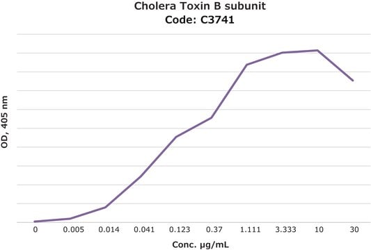
Activity measured by ELISA using ganglioside GM1-coated multiwell plates, rabbit anti-Cholera toxin B subunit, and peroxidase-labeled goat anti-rabbit IgG as the secondary antibody. 50% saturation of binding is achieved with 0.02-1 μg of Cholera toxin B subunit-biotin conjugate per mL. The conjugated B subunit gives a similar value for 50% binding to that of unconjugated B subunit from which it is prepared.

Hazard Statements

Precautionary Statements

Hazard Classifications

Aquatic Chronic 3

Storage Class Code

11 - Combustible Solids

WGK

WGK 3

Flash Point(F)

Not applicable

Flash Point(C)

Not applicable

Personal Protective Equipment

dust mask type N95 (US), Eyeshields, Gloves

Choose from one of the most recent versions:

Certificates of Analysis (COA)

Lot/Batch Number

Don't see the Right Version?

If you require a particular version, you can look up a specific certificate by the Lot or Batch number.

Already Own This Product?

Find documentation for the products that you have recently purchased in the Document Library.

Visit the Document Library

Evidence for TLR4 and FcRgamma-CARD9 activation by cholera toxin B subunit and its direct bindings to TREM2 and LMIR5 receptors
Phongsisay V, et al.
Molecular Immunology, 66(2), 463-471 (2015)
The Ia. 2 epitope defines a subset of lipid raft-resident MHC class II molecules crucial to effective antigen presentation
Busman-Sahay K, et al.
Journal of Immunology, 1100336-1100336 (2011)
Lucia Gardini et al.
Scientific reports, 5, 16088-16088 (2015-11-04)
Live cells are three-dimensional environments where biological molecules move to find their targets and accomplish their functions. However, up to now, most single molecule investigations have been limited to bi-dimensional studies owing to the complexity of 3d-tracking techniques. Here, we
3D tracking of single nanoparticles and quantum dots in living cells by out-of-focus imaging with diffraction pattern recognition
Gardini L, et al.
Scientific Reports, 5, 16088-16088 (2015)
Eva Koffeman et al.
Methods in molecular medicine, 136, 69-86 (2007-11-07)
T-cells specific for a particular antigen represent a small percentage of the overall T-cell population. Detecting the presence of antigen specific T-cells in patients, animal models or populations of cultured cells has presented a challenge to researchers. The T-cell capture

Our team of scientists has experience in all areas of research including Life Science, Material Science, Chemical Synthesis, Chromatography, Analytical and many others.

Contact Technical Service