Skip to Content
Merck
All Photos(2)

Key Documents

D8168

Sigma-Aldrich

Anti-Dystrophin Antibody

mouse monoclonal, MANDYS8

Synonym(s):

Dystrophin Antibody, Dystrophin Antibody - Monoclonal Anti-Dystrophin antibody produced in mouse

Sign Into View Organizational & Contract Pricing


About This Item

MDL number:
UNSPSC Code:
12352203
NACRES:
NA.41

Product Name

Monoclonal Anti-Dystrophin antibody produced in mouse, clone MANDYS8, ascites fluid

biological source

mouse

Quality Level

conjugate

unconjugated

antibody form

ascites fluid

antibody product type

primary antibodies

clone

MANDYS8, monoclonal

species reactivity

chicken, rat, human, pig, rabbit, mouse

technique(s)

indirect ELISA: suitable
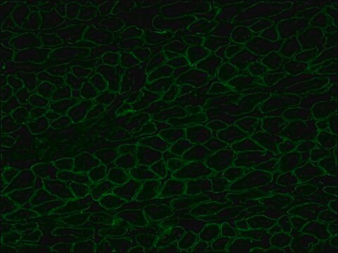
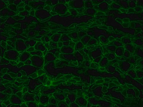
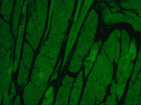
indirect immunofluorescence: 1:400 using frozen human or animal muscle tissue sections.
microarray: suitable
western blot: suitable

isotype

IgG2b

UniProt accession no.

shipped in

dry ice

storage temp.

−20°C

target post-translational modification

unmodified

Gene Information

human ... DMD(1756)
mouse ... Dmd(13405)
rat ... Dmd(24907)

General description

Monoclonal Anti-Dystrophin (mouse IgG2b isotype) is derived from the MANDYS8 hybridoma produced by the fusion of mouse myeloma cells and splenocytes from an immunized mouse. Dystrophin is a structural protein on the inner face of the membrane, consisting of a 25-repeat, rod-like, triple-helical domain separating an N-terminal actin binding domain from two C-terminal domains, one of which is rich in cysteine.

Immunogen

recombinant human dystrophin fragment.

Application

Monoclonal Anti-Dystrophin antibody produced in mouse has been used in
  • immunohistochemistry
  • immunofluorescence
  • double immunofluorescence terminal dUTP nick-end labeling (TUNEL)
  • immunoblotting

Monoclonal anti-dystrophin antibody can be used for localization of dystrophin using immunochemical assays like ELISA and immunohistochemistry. The antibody can also be used in immunoblotting for brain dystrophin. Further, it can be used in western blotting and double immunofluorescent labelling (diluted 1: 500) of dystrophin.

Biochem/physiol Actions

Dystrophin deficiency is associated with severe Duchenne muscular dystrophy (DMD). Becker muscular dystrophy (BMD) show less pronounced abnormalities of dystrophin protein expression. Since abnormalities in the protein expression occur specifically in patients with these types of muscular dystrophy, dystrophin analysis may be used to distinguish these conditions from other neuromuscular diseases.

Target description

The rod domain of the human dystrophin molecule is present in normal muscle tissue and in nearly all Becker muscular dystrophies. It is absent in the cases of Duchenne muscular dystrophies and in the dystrophic mouse (mdx).

Other Notes

This product can be found as purified product that was produced using cell culture hybridoma product.
SAB4200764 Anti-Dystrophin antibody, Mouse monoclonal
clone MANDYS8, purified from hybridoma cell culture

Disclaimer

Unless otherwise stated in our catalog or other company documentation accompanying the product(s), our products are intended for research use only and are not to be used for any other purpose, which includes but is not limited to, unauthorized commercial uses, in vitro diagnostic uses, ex vivo or in vivo therapeutic uses or any type of consumption or application to humans or animals.

Not finding the right product?  

Try our Product Selector Tool.

Storage Class Code

10 - Combustible liquids

WGK

WGK 3

Flash Point(F)

Not applicable

Flash Point(C)

Not applicable


Choose from one of the most recent versions:

Certificates of Analysis (COA)

Lot/Batch Number

Don't see the Right Version?

If you require a particular version, you can look up a specific certificate by the Lot or Batch number.

Already Own This Product?

Find documentation for the products that you have recently purchased in the Document Library.

Visit the Document Library

The dystrophin complex: structure, function, and implications for therapy
Gao QQ and McNally EM
Comprehensive Physiology, 5(3), 1223-1239 (2011)
Rachel Blitzblau et al.
Brain research, 1218, 21-34 (2008-06-06)
Muscular dystrophy patients often show cognitive impairment, in addition to muscle degeneration caused by dystrophin gene defects. The cognitive impairments lead to speculation that the dystrophin protein family may play a key role at neuronal synapses. Dystrophin regulates the stability
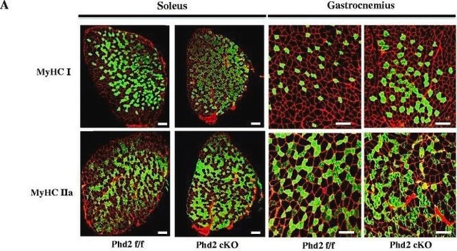
Myospryn is a calcineurin-interacting protein that negatively modulates slow-fiber-type transformation and skeletal muscle regeneration
Kielbasa OM, et al.
Faseb Journal, 25(7), 2276-2286 (2011)
Effects of long-term resveratrol-induced SIRT1 activation on insulin and apoptotic signalling in aged skeletal muscle
Sin TK, et al.
Acta Diabetologica, 52(6), 1063-1075 (2015)
Juliette A Strauss et al.
Physiological reports, 4(1) (2016-01-07)
Synaptosomal-associated protein 23 (SNAP23) is a SNARE protein expressed abundantly in human skeletal muscle. Its established role is to mediate insulin-stimulated docking and fusion of glucose transporter 4 (GLUT4) with the plasma membrane. Recent in vitro research has proposed that SNAP23

Our team of scientists has experience in all areas of research including Life Science, Material Science, Chemical Synthesis, Chromatography, Analytical and many others.

Contact Technical Service