Skip to Content
Merck
All Photos(2)

Documents

A2429

Sigma-Aldrich

Anti-Mouse IgG (Fc specific)–Alkaline Phosphatase antibody produced in goat

affinity isolated antibody, buffered aqueous solution

Synonym(s):

Goat Anti-Mouse IgG (Fc specific)–AP

Sign Into View Organizational & Contract Pricing


About This Item

MDL number:
UNSPSC Code:
12352203
NACRES:
NA.46

biological source

goat

conjugate

alkaline phosphatase conjugate

antibody form

affinity isolated antibody

antibody product type

secondary antibodies

clone

polyclonal

form

buffered aqueous solution

species reactivity

mouse

should not react with

bovine, human, horse

technique(s)

direct ELISA: 1:30,000
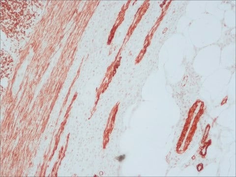
immunohistochemistry (formalin-fixed, paraffin-embedded sections): 1:40
western blot (chemiluminescent): 1:160,000-1:320,000

shipped in

wet ice

storage temp.

2-8°C

target post-translational modification

unmodified

Looking for similar products? Visit Product Comparison Guide

General description

Immunoglobulin G (IgG) belongs to the immunoglobulin family and is a widely expressed serum antibody. An immunoglobulin has two heavy chains and two light chains connected by a disulfide bond. It is a glycoprotein. IgG is a major class of immunoglobulin. Mouse consists of five immunoglobulin classes- IgM, IgG, IgA, IgD and IgE. Mouse IgG is further divided into five classes- IgG1, IgG2a, IgG2b and IgG3.
Immunoglobulin G (IgG) is a glycoprotein antibody that regulates immune responses such as phagocytosis and is also involved in the development of autoimmune diseases. IgG1 regulates complement fixation in mice.
Mouse IgG is a plasma B cell derived antibody isotype defined by its heavy chain. IgG is the most abundant antibody isotype found in mouse serum. IgG crosses the placental barrier, is a complement activator and binds to the Fc-receptors on phagocytic cells. The level of IgG may vary with the status of disease or infection.

Immunogen

Purified mouse IgG, Fc fragment

Application

Alkaline phosphatase-conjugated goat anti-mouse Fc specific antibody was used as a secondary antibody in ELISA assays at a dilution of 1:1000 in PBS/0.1% Tween and 1% BSA for 1.5 hours at 37°C. Antibody was developed using 4-nitrophenyl phosphate (Sigma) as a substrate for 30 minutes at 37°C.
Anti-Mouse IgG (Fc specific)-Alkaline Phosphatase antibody is suitable for use in ELISA (1:1500) and western blot. The antibody can also be used for immunohistochemistry (1:40 using formalin-fixed, paraffin-embedded sections).
Anti-Mouse IgG (Fc specific)-Alkaline Phosphatase antibody produced in goat has been used in enzyme-linked immunosorbent assay (ELISA), sandwich ELISA and enzyme-linked immune absorbent spot (ELISPOT).
The product has been used as a secondary antibody for detecting mouse serum anti-hemagglutinin (HA) antibodies using ELISA techniques.

Biochem/physiol Actions

Immunoglobulin G (IgG) participates in hypersensitivity type II and type III reactions. IgG helps in opsonization, complement fixation and antibody dependent cell mediated cytotoxicity.

Other Notes

Antibody adsorbed with bovine, equine and human serum proteins.

Physical form

Solution in 0.05 M Tris, pH 8.0, containing 1% bovine serum albumin, 1 mM MgCl2 and 15 mM sodium azide.

Preparation Note

Adsorbed to reduce background staining with bovine, horse, or human samples.

Disclaimer

Unless otherwise stated in our catalog or other company documentation accompanying the product(s), our products are intended for research use only and are not to be used for any other purpose, which includes but is not limited to, unauthorized commercial uses, in vitro diagnostic uses, ex vivo or in vivo therapeutic uses or any type of consumption or application to humans or animals.

Not finding the right product?  

Try our Product Selector Tool.

Storage Class Code

10 - Combustible liquids

WGK

WGK 1

Flash Point(F)

Not applicable

Flash Point(C)

Not applicable


Certificates of Analysis (COA)

Search for Certificates of Analysis (COA) by entering the products Lot/Batch Number. Lot and Batch Numbers can be found on a product’s label following the words ‘Lot’ or ‘Batch’.

Already Own This Product?

Find documentation for the products that you have recently purchased in the Document Library.

Visit the Document Library

Arnar Gudjonsson et al.
Journal of immunology (Baltimore, Md. : 1950), 198(7), 2785-2795 (2017-02-24)
Targeting Ags to conventional dendritic cells can enhance Ag-specific immune responses. Although most studies have focused on the induction of T cell responses, the mechanisms by which targeting improves Ab responses are poorly understood. In this study we present data
Xiaogang Wang et al.
Nature communications, 9(1), 1379-1379 (2018-04-13)
Secretion of extracellular vesicles (EVs), a process common to eukaryotes, archae, and bacteria, represents a secretory pathway that allows cell-free intercellular communication. Microbial EVs package diverse proteins and influence the host-pathogen interaction, but the mechanisms underlying EV production in Gram-positive
Pedro Humberto Castro et al.
Journal of experimental botany, 69(19), 4633-4649 (2018-07-28)
Post-translational modifiers such as the small ubiquitin-like modifier (SUMO) peptide act as fast and reversible protein regulators. Functional characterization of the sumoylation machinery has determined the key regulatory role that SUMO plays in plant development. Unlike components of the SUMO
Marcel Zocher et al.
International immunology, 15(7), 789-796 (2003-06-17)
Antigen-specific B cells are key players in many autoimmune diseases through the production of autoreactive antibodies that can cause severe tissue damage and malfunction. We have designed and expressed a fusion protein, referred to as MOG-Fc, composed of the extracellular
The specificity of targeted vaccines for APC surface molecules influences the immune response phenotype
Grodeland G, et al.
Testing, 8(11), e80008-e80008 (2013)

Our team of scientists has experience in all areas of research including Life Science, Material Science, Chemical Synthesis, Chromatography, Analytical and many others.

Contact Technical Service