General description
Human C-terminal-binding protein 2 (CtBP2) and Ribeye (UniProt P56545-1 and P56545-2, respectively) are two proteins encoded by the CTBP2 gene (Gene ID 1488) in human, they are produced as a result of alternative splicing. The C-terminal 425-amino acid sequence of Ribeye (aa 561-985) is identical to that of CtBP2 (aa 21-445), however the first 20 amino acids at the CtBP2 N-terminal end is replaced with a 560-amino acid sequence in the case of Ribeye. Ribeye is a protein specific to synaptic ribbons. Ribbon synapses are high-performance synapses built by certain nonspiking sensory neurons, such as photoreceptors, retinal bipolar cells, and inner ear hair cells. The main site of exocytosis of these neurons is characterized by unique presynaptic structures called the synaptic ribbons that associate with large numbers of synaptic vesicles primed for exocytosis and has a direct impact on synaptic signaling. Ribeye is composed of an N-terminal A domain and a C-terminal B domain that is largely identical to CtBP2. The A domain unique to Ribeye has a structural role at the synaptic ribbon, while the B domain of Ribeye possesses lysophosphatidic acid (LPA) acyltransferase (LPAAT) activity that catalyzes the generation of the highly active signaling phospholipid phosphatidic acid (PA) from LPA. LPAAT-deficient Ribeye mutant does not recruit PA-binding proteins to synaptic ribbons, whereas wild-type RIBEYE supports PA-dependent binding, The B domain also contains an NADH-binding subdomain (NBD), which is reported to compete against Arf1 for Arf-GTPase activating protein-3 (ArfGAP3) binding. Ribeye therefore functions as an Arf1 positive regulator at the synaptic ribbon by preventing the inactivation of Arf1 by ArfGAP3.
Specificity
Specific for CTBP2 spliced variant Ribeye. Does not react with the canonical CtBP2 isoform.
Immunogen
Epitope: N-terminal region
GST-tagged recombinant protein corresponding to the N-terminal region of human Ribeye.
Application
Anti-Ribeye Antibody, clone 3D12.1, Cat. No. MABN804, is a highly specific mouse monoclonal antibody, that targets Ribeye and has been tested in Western Blotting and Immunohistochemistry (Paraffin).
Research Category
Neuroscience
Research Sub Category
Developmental Neuroscience
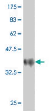
Western Blotting Analysis: 0.5 µg/mL from a representative lot detected Ribeye in 10 µg of SH-SY5Y cell lysate.
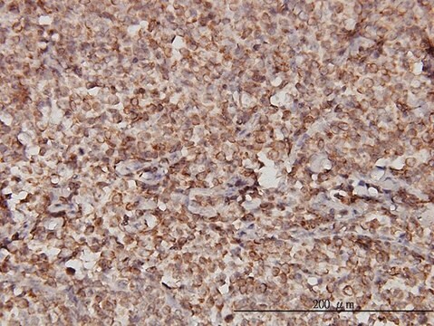
Immunohistochemistry Analysis: A 1:50-250 dilution from a representative lot detected Ribeye in human cardiac myocytes and human cerebellum tissues.
Quality
Evaluated by Western Blotting in HeLa cell lysate.
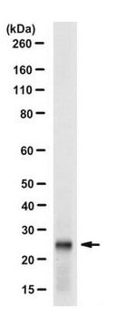
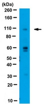
Western Blotting Analysis: 0.5 µg/mL of this antibody detected Ribeye in 10 µg of HeLa cell lysate.
Target description
~106 kDa observed (109.2 kDa calculated).
Physical form
Format: Purified
Protein G Purified
Purified mouse monoclonal IgG1κ antibody in buffer containing 0.1 M Tris-Glycine (pH 7.4), 150 mM NaCl with 0.05% sodium azide.
Storage and Stability
Stable for 1 year at 2-8°C from date of receipt.
Other Notes
Concentration: Please refer to lot specific datasheet.
Disclaimer
Unless otherwise stated in our catalog or other company documentation accompanying the product(s), our products are intended for research use only and are not to be used for any other purpose, which includes but is not limited to, unauthorized commercial uses, in vitro diagnostic uses, ex vivo or in vivo therapeutic uses or any type of consumption or application to humans or animals.
