Skip to Content
Merck
All Photos(1)

Documents

S9442

Sigma-Aldrich

Anti-Synaptopodin (SE-19) antibody produced in rabbit

IgG fraction of antiserum, buffered aqueous solution

Sign Into View Organizational & Contract Pricing


About This Item

MDL number:
UNSPSC Code:
12352203
NACRES:
NA.41

biological source

rabbit

conjugate

unconjugated

antibody form

IgG fraction of antiserum

antibody product type

primary antibodies

clone

polyclonal

form

buffered aqueous solution

mol wt

antigen 100 kDa

species reactivity

rat

technique(s)

microarray: suitable
western blot: 1:3,000 using cytosolic S1 fraction of rat brain

UniProt accession no.

shipped in

dry ice

storage temp.

−20°C

target post-translational modification

unmodified

Gene Information

human ... SYNPO(11346)
rat ... Synpo(60324)

Related Categories

General description

Synaptopodin is a proline-rich, actin-associated protein (100 kDa) is present in the post-synaptic density (PSD) in actin cytoskeleton. It is also present in the dendritic spines and differentiated podocytes. Synaptopodin contains a high amount of proline residues (~20%), which are evenly distributed along the protein sequence, thus virtually excluding the formation of globular domains. In particular, synaptopodin does not contain functional domains found in receptor clustering PSD proteins. Sequence comparison between human and mouse synaptopodin shows 84% identity. The exclusive site of synaptopodin expression in the telencephalon is restricted to specific areas of high synaptic plasticity, i.e. in the perykaria of the olfactory bulb, striatum, and hippocampus.

Immunogen

synthetic peptide corresponding to amino acids 184-202 of rat synaptopodin conjugated to KLH. This rat synaptopodin sequence has ~70% identity with human synaptopodin.

Application

Anti-Synaptopodin (SE-19) antibody produced in rabbit has been used in:
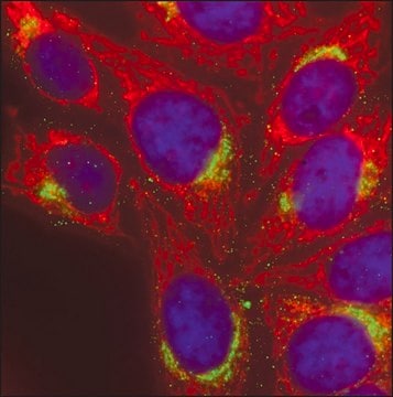
  • immunofluorescence microscopy
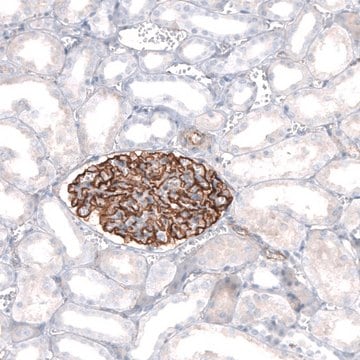
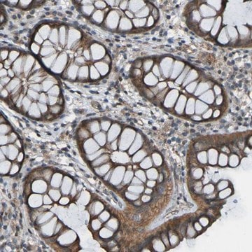
  • immunohistochemistry
  • western blotting

Biochem/physiol Actions

Synaptopodin is thought to play an important role in synaptic plasticity and is associated in the modulation of motility of actin in neuronal dendritic spines. The association of synaptopodin with the specialized actin system of renal podocyte foot processes suggests that it may play a role in actin cytoskeleton dynamics of these cellular extensions. Synaptopodin has been found to interact with α-actinin-4 and the tight junction protein Membrane-associated guanylate kinase inverted 1 (MAGI-1) in Madin-Darby Canine Kidney (MDCK) cells.

Physical form

Solution in 0.01 M phosphate buffered saline, pH 7.4, containing 15 mM sodium azide.

Disclaimer

Unless otherwise stated in our catalog or other company documentation accompanying the product(s), our products are intended for research use only and are not to be used for any other purpose, which includes but is not limited to, unauthorized commercial uses, in vitro diagnostic uses, ex vivo or in vivo therapeutic uses or any type of consumption or application to humans or animals.

Not finding the right product?  

Try our Product Selector Tool.

Pictograms

Exclamation mark

Signal Word

Warning

Hazard Statements

Precautionary Statements

Hazard Classifications

Acute Tox. 4 Oral - Aquatic Chronic 3

Storage Class Code

10 - Combustible liquids

WGK

WGK 3

Flash Point(F)

Not applicable

Flash Point(C)

Not applicable


Certificates of Analysis (COA)

Search for Certificates of Analysis (COA) by entering the products Lot/Batch Number. Lot and Batch Numbers can be found on a product’s label following the words ‘Lot’ or ‘Batch’.

Already Own This Product?

Find documentation for the products that you have recently purchased in the Document Library.

Visit the Document Library

Synaptopodin is regulated by aromatase activity
Fester L, et al.
Journal of Neurochemistry, 140(1), 126-139 (2017)
Synaptopodin: an actin-associated protein in telencephalic dendrites and renal podocytes
Mundel P, et al.
The Journal of cell biology, 139(1), 193-204 (1997)
Expression of synaptopodin, an actin-associated protein, in the rat hippocampus after limbic epilepsy
Roth SU, et al.
Brain Pathology, 11(2), 169-181 (2001)
Alberto Perez-Alvarez et al.
Nature communications, 11(1), 5083-5083 (2020-10-10)
In hippocampal pyramidal cells, a small subset of dendritic spines contain endoplasmic reticulum (ER). In large spines, ER frequently forms a spine apparatus, while smaller spines contain just a single tubule of smooth ER. Here we show that the ER
A dual agonist of farnesoid X receptor (FXR) and the G protein-coupled receptor TGR5, INT-767, reverses age-related kidney disease in mice
Wang XX, et al.
Test, 292(29), 12018-12024 (2017)

Our team of scientists has experience in all areas of research including Life Science, Material Science, Chemical Synthesis, Chromatography, Analytical and many others.

Contact Technical Service