Skip to Content
Merck
All Photos(1)

Key Documents

D8156

Sigma-Aldrich

Monoclonal Anti-Digoxin antibody produced in mouse

clone DI-22, ascites fluid

Synonym(s):

Monoclonal Anti-Digoxin

Sign Into View Organizational & Contract Pricing


About This Item

UNSPSC Code:
12352203
NACRES:
NA.43

biological source

mouse

Quality Level

conjugate

unconjugated

antibody form

ascites fluid

antibody product type

primary antibodies

clone

DI-22, monoclonal

contains

15 mM sodium azide

technique(s)

dot blot: suitable
flow cytometry: suitable
indirect ELISA: 1:10,000 using digoxin-BSA
indirect ELISA: 1:2,500 using digoxigenin-transferrin

isotype

IgG1

shipped in

dry ice

storage temp.

−20°C

target post-translational modification

unmodified

Looking for similar products? Visit Product Comparison Guide

General description

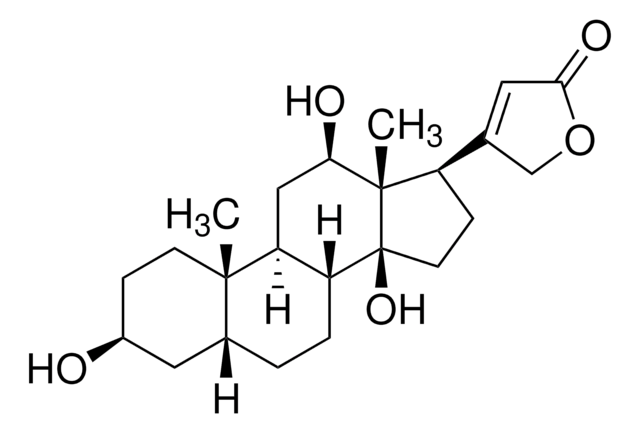
Monoclonal Anti-Digoxin (mouse IgG1 isotype) is derived from the hybridoma DI-22 produced by the fusion of mouse myeloma cells and splenocytes from a BALB/c mouse immunized with digoxin- keyhole limpet hemocyanin (KLH). Digoxin is obatained from the leaves of the Digitalis lanata, a foxglove plant. The digoxin molecule is made up of a sugar and a cardenolide. It has a molecular weight is 780.95 Da. It is a odorless white crystal, which is soluble in alcohol and freely soluble in pyridine. It is expressed in the duodenum, kidneys, liver and the blood-brain barrier.

Specificity

Antibody is specific for digoxin and shows a high affinity for digoxigenin.
Monoclonal Anti-Digoxin antibody is specific for digoxin and digoxin-labeled compounds, and shows strong cross-reactivity with digoxigenin.

Immunogen

Digoxin-KLH.

Application

Monoclonal Anti-Digoxin antibody produced in mouse has been used in:
  • enzyme-linked immunosorbent assay (ELISA)
  • dot blot
  • flow cytometry
  • fluorescence in situ hybridization (FISH)
  • DNA hybridization
  • in-situ hybridization (ISH)
  • immunodetection of the probe and chromosome X

Biochem/physiol Actions

Digoxin is a phytoestrogen that binds estrogen receptors and produces inotropic effects in cardiac muscle. Studies show that digoxin decreases the risk of breast and uterus cancers that are estrogen sensitive.
Digoxin is a substrate for P-glycoprotein (P-gp), a membrane efflux transporter. This antibody may be used to detect digoxin-labeled compounds such as oligonucleotides, antibodies or peptides.

Disclaimer

Unless otherwise stated in our catalog or other company documentation accompanying the product(s), our products are intended for research use only and are not to be used for any other purpose, which includes but is not limited to, unauthorized commercial uses, in vitro diagnostic uses, ex vivo or in vivo therapeutic uses or any type of consumption or application to humans or animals.

Not finding the right product?  

Try our Product Selector Tool.

Storage Class Code

12 - Non Combustible Liquids

WGK

nwg

Flash Point(F)

Not applicable

Flash Point(C)

Not applicable


Certificates of Analysis (COA)

Search for Certificates of Analysis (COA) by entering the products Lot/Batch Number. Lot and Batch Numbers can be found on a product’s label following the words ‘Lot’ or ‘Batch’.

Already Own This Product?

Find documentation for the products that you have recently purchased in the Document Library.

Visit the Document Library

Drug Metabolism in Cardiovascular Disease
Drug Metabolism in Diseases, 139-156 (2017)
Effect of hormones on the concentration of a digoxin-like material in Tetrahymena
Csaba G, et al.
Acta Protozoologica, 44, 81-84 (2005)
Digoxin
Small Animal Clinical Pharmacology, 805-806 (2009)
Ying-Liang Duan et al.
Journal of virology, 88(7), 3861-3873 (2014-01-24)
After infection, human cytomegalovirus (HCMV) persists for life. Primary infections and reactivation of latent virus can both result in congenital infection, a leading cause of central nervous system birth defects. We previously reported long-term HCMV infection in the T98G glioblastoma
Waqar Ahmed et al.
Scientific reports, 8(1), 15438-15438 (2018-10-20)
Epstein-Barr virus-encoded RNAs (EBER1 and EBER2) are two highly abundant, non-protein coding RNAs consistently expressed in all EBV infected cells, but their function remains poorly understood. Conventional in situ hybridization studies have indicated that these RNAs are present exclusively in

Our team of scientists has experience in all areas of research including Life Science, Material Science, Chemical Synthesis, Chromatography, Analytical and many others.

Contact Technical Service