Skip to Content
Merck
All Photos(2)

Documents

T2780

Sigma-Aldrich

Monoclonal Anti-Tropomyosin antibody produced in mouse

clone TM311, ascites fluid

Synonym(s):

Anti-Tropomyosin Antibody

Sign Into View Organizational & Contract Pricing


About This Item

MDL number:
UNSPSC Code:
12352203
NACRES:
NA.41

biological source

mouse

Quality Level

conjugate

unconjugated

antibody form

ascites fluid

antibody product type

primary antibodies

clone

TM311, monoclonal

mol wt

antigen 36-39 kDa

contains

15 mM sodium azide

species reactivity

pig, chicken, bovine, rabbit, mouse, hamster, rat, human

technique(s)

immunoprecipitation (IP): suitable
indirect ELISA: suitable
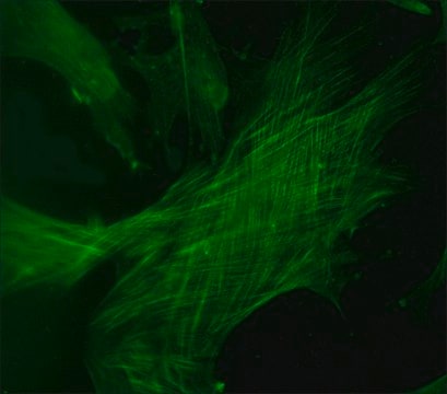
indirect immunofluorescence: 1:400 using chicken fibroblasts
microarray: suitable
western blot: suitable using human tissue and chicken gizzard extracts

isotype

IgG1

UniProt accession no.

shipped in

dry ice

storage temp.

−20°C

target post-translational modification

unmodified

Gene Information

bovine ... TPM1(281544)
chicken ... TPM1(396366)
human ... TPM1(7168)
mouse ... Tpm1(22003)
rat ... Tpm1(24851)

General description

Monoclonal Anti-Tropomyosin (mouse IgG1 isotype) is derived from the hybridoma produced by the fusion of mouse myeloma cells and splenocytes from an immunized mouse. Tropomyosin is a rigid rod-shaped protein closely associated with actin filaments. Non-muscle forms of tropomyosin have been identified in a wide range of cell types. Tropomyosin is made of two α helical polypeptide chains.

Immunogen

chicken gizzard tropomyosin.

Application

Monoclonal Anti-Tropomyosin has been used:
  • in immunofluorescent labelling
  • in immunohistochemistry
  • in western blotting
  • in immunoblotting

Mouse monoclonal clone TM311 anti-Tropomyosin antibody is used to tag tropomyosin for detection and quantitation by immunocytochemical and immunohistochemical (IHC) techniques such as immunoblotting, immunoprecipitation, and immunofluorescence. It is used as a probe to determine the presence and roles of tropomyosin in cytoskeletal structures.

Biochem/physiol Actions

Tropomyosin together with troponin, regulate the binding of myosin to actin. Tropomyosin is a dimeric coiled-coil protein forming continuous polymers along the myosin-binding groove of actin. Various tropomyosin isoforms help to confer structure and function to actins in the cytoskeleton and in striated muscle function.

Disclaimer

Unless otherwise stated in our catalog or other company documentation accompanying the product(s), our products are intended for research use only and are not to be used for any other purpose, which includes but is not limited to, unauthorized commercial uses, in vitro diagnostic uses, ex vivo or in vivo therapeutic uses or any type of consumption or application to humans or animals.

Not finding the right product?  

Try our Product Selector Tool.

Storage Class Code

10 - Combustible liquids

WGK

nwg

Flash Point(F)

Not applicable

Flash Point(C)

Not applicable


Certificates of Analysis (COA)

Search for Certificates of Analysis (COA) by entering the products Lot/Batch Number. Lot and Batch Numbers can be found on a product’s label following the words ‘Lot’ or ‘Batch’.

Already Own This Product?

Find documentation for the products that you have recently purchased in the Document Library.

Visit the Document Library

Activation of MAP kinases and phosphorylation of caldesmon in canine colonic smooth muscle.
Gerthoffer W T, et al.
The Journal of Physiology, 495(3), 597-609 (1996)
Ewelina Jurewicz et al.
Biochimica et biophysica acta, 1833(3), 761-766 (2012-12-26)
The CacyBP/SIP protein interacts with several targets, including actin. Since the majority of actin filaments are associated with tropomyosin, in this work we characterized binding of CacyBP/SIP to the actin-tropomyosin complex and examined the effects of CacyBP/SIP on actin filament
Tropomyosin-master regulator of actin filament function in the cytoskeleton
Gunning P W, et al.
Journal of Cell Science, 128(16), 2965-2974 (2015)
CENP-A is essential for cardiac progenitor cell proliferation
McGregor M, et al
Cell Cycle, 13(5), 739-748 (2014)
Non-canonical Wnt signaling enhances differentiation of Sca1+/c-kit+ adipose-derived murine stromal vascular cells into spontaneously beating cardiac myocytes
Palpant N J, et al.
Journal of Molecular and Cellular Cardiology, 43(3), 362-370 (2007)

Our team of scientists has experience in all areas of research including Life Science, Material Science, Chemical Synthesis, Chromatography, Analytical and many others.

Contact Technical Service