Skip to Content
Merck
All Photos(2)

Key Documents

ABC232

Sigma-Aldrich

Anti-MAP1LC3A antibody

rabbit polyclonal

Synonym(s):

Microtubule-associated proteins 1A/1B light chain 3A, Autophagy-related protein LC3 A, Autophagy-related ubiquitin-like modifier LC3 A, MAP1 light chain 3-like protein 1, MAP1A/MAP1B light chain 3 A, MAP1A/MAP1B LC3 A, Microtubule-associated protein 1 li

Sign Into View Organizational & Contract Pricing


About This Item

UNSPSC Code:
12352203
eCl@ss:
32160702
NACRES:
NA.41

Product Name

Anti-LC3 Antibody, serum, from rabbit

biological source

rabbit

Quality Level

antibody form

serum

antibody product type

primary antibodies

clone

polyclonal

species reactivity

human, rat, mouse

technique(s)

western blot: suitable

NCBI accession no.

UniProt accession no.

shipped in

wet ice

target post-translational modification

unmodified

Gene Information

General description

LC3A is composed of a heavy chain and multiple light-chain subunits. The molecular machinery required for autophagy is highly conserved in all eukaryotes as seen by the high degree of conservation of proteins involved in the formation of the autophagosome membranes. Recently, both yeast Apg8p and its rat homologue LC3-II were identified as essential constituents of autophagosome membrane as a processed form. In addition, both the yeast and human proteins exist in two modified forms produced by a series of post-translational modifications including a critical C-terminal cleavage after a conserved Gly residue, and the smaller processed form is associated with the autophagosome membranes Results revealed different regulation of the three human isoforms of LC3-II and implicate that the three isoforms may have different physiological functions. Two forms of LC3A, called LC3-I and -II, were produced post-translationally in various cells. LC3-I is cytosolic, whereas LC3-II is membrane bound.

Immunogen

Linear peptide corresponding to human LC3.

Application

Anti-LC3 Antibody is an antibody against LC3 for use in Western Blotting.
Research Category
Apoptosis & Cancer
Research Sub Category
Apoptosis - Additional
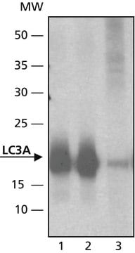
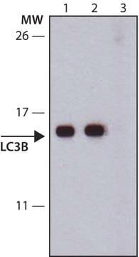
Western Blot Analysis: A 1;1,000 dilution from a representative lot detected LC3 in 10 µg of mouse embryonic fibroblast tissue lysate and MCF-7 cell lysate.

Quality

Evaluated by Western Blot in NIH/3T3 cell lysate.

Western Blot Analysis: A 1;1,000 dilution of this antibody detected LC3 in 10 µg of NIH/3T3 cell lysate.

Target description

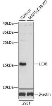
~16 kDa observed. LC3 can occur as a doublet around ~15 kDa, indicating the presence of both LC3-I & LC3-II. An uncharacterized band may appear at ~39 and ~55 kDa in some lysates.

Linkage

Replaces: MABC177; MABC176

Physical form

Rabbit polyclonal serum containing 0.05% sodium azide.
Unpurified

Storage and Stability

Stable for 1 year at -20°C from date of receipt.
Handling Recommendations: Upon receipt and prior to removing the cap, centrifuge the vial and gently mix the solution. Aliquot into microcentrifuge tubes and store at -20°C. Avoid repeated freeze/thaw cycles, which may damage IgG and affect product performance.

Analysis Note

Control
NIH/3T3 cell lysate

Disclaimer

Unless otherwise stated in our catalog or other company documentation accompanying the product(s), our products are intended for research use only and are not to be used for any other purpose, which includes but is not limited to, unauthorized commercial uses, in vitro diagnostic uses, ex vivo or in vivo therapeutic uses or any type of consumption or application to humans or animals.

Not finding the right product?  

Try our Product Selector Tool.

Storage Class Code

10 - Combustible liquids

WGK

WGK 1


Certificates of Analysis (COA)

Search for Certificates of Analysis (COA) by entering the products Lot/Batch Number. Lot and Batch Numbers can be found on a product’s label following the words ‘Lot’ or ‘Batch’.

Already Own This Product?

Find documentation for the products that you have recently purchased in the Document Library.

Visit the Document Library

Ribosomal protein mutations induce autophagy through S6 kinase inhibition of the insulin pathway.
Heijnen, HF; van Wijk, R; Pereboom, TC; Goos, YJ; Seinen, CW; van Oirschot et al.
PLoS Genetics null
Rosalba Siracusa et al.
International journal of molecular sciences, 22(10) (2021-06-03)
Endometriosis is a gynecological condition affecting patients in reproductive age. The aim of this paper was to assess the effects of the autophagy and mitophagy induction in a rat model of endometriosis. Endometriosis was induced by the injection of uterine
Rui Kong
Cell biology international, 44(9), 1945-1956 (2020-05-26)
Non-small-cell lung carcinoma (NSCLC) continues to top the list of cancer mortalities worldwide. The role of circular RNAs (circRNAs) in tumorigenesis has been increasingly appreciated, although it is relatively unexplored in NSCLC. Herein, we reported the role of hsa_circ_0085131 in
Florie A Mar et al.
Autophagy, 11(3), 527-537 (2015-03-10)
Telomere dysfunction plays a complex role in tumorigenesis. While dysfunctional telomeres can block the proliferation of incipient cancer clones by inducing replicative senescence, fusion of dysfunctional telomeres can drive genome instability and oncogenic genomic rearrangements. Therefore, it is important to
Aleksandra V Borodkina et al.
Cell cycle (Georgetown, Tex.), 15(1), 117-127 (2015-12-05)
Previously we demonstrated that endometrium-derived human mesenchymal stem cells (hMESCs) via activation of the ATM/p53/p21/Rb pathway enter the premature senescence in response to oxidative stress. Down regulation effects of the key components of this signaling pathway, particularly ATM and p53

Articles

Autophagy is a regulated process involved in cell growth, development, and recycling of cytoplasmic components in cells.

Autophagy is a regulated process involved in cell growth, development, and recycling of cytoplasmic components in cells.

Autophagy is a regulated process involved in cell growth, development, and recycling of cytoplasmic components in cells.

Autophagy is a regulated process involved in cell growth, development, and recycling of cytoplasmic components in cells.

Our team of scientists has experience in all areas of research including Life Science, Material Science, Chemical Synthesis, Chromatography, Analytical and many others.

Contact Technical Service