AB5895
Anti-P2X3 Receptor Antibody, pain
serum, Chemicon®
Sign Into View Organizational & Contract Pricing
All Photos(1)
About This Item
UNSPSC Code:
12352203
eCl@ss:
32160702
NACRES:
NA.41
Recommended Products
biological source
rabbit
Quality Level
antibody form
serum
antibody product type
primary antibodies
clone
polyclonal
species reactivity
rat, human, primate
manufacturer/tradename
Chemicon®
technique(s)
immunocytochemistry: suitable
immunohistochemistry: suitable
western blot: suitable
NCBI accession no.
UniProt accession no.
shipped in
dry ice
target post-translational modification
unmodified
Gene Information
human ... P2RX3(5024)
Specificity
P2X3
SPECIES REACTIVITIES:
Rat, mouse and primate. Other species have not been tested.
SPECIES REACTIVITIES:
Rat, mouse and primate. Other species have not been tested.
Immunogen
Synthetic peptide corresponding to the C-terminus of rat P2X3.
Application
Detect P2X3 Receptor using this Anti-P2X3 Receptor Antibody, pain validated for use in IC, IH & WB.
Immunoblotting: 1:500-1:1,000
Immunohistochemistry: 1:500-1:1,000
Immunocytochemistry: 1:500-1:1,000
Optimal working dilutions must be determined by the end user.
APPLICATION NOTES FOR AB5895
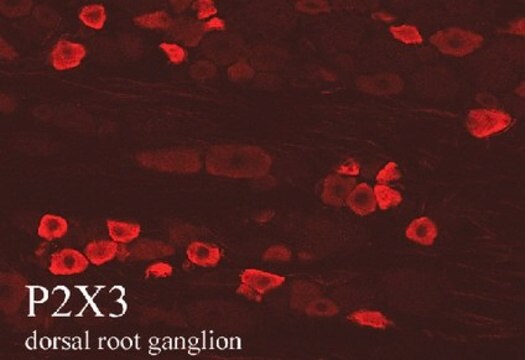
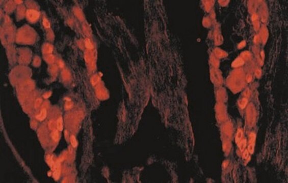
IMMUNOHISTOCHEMISTRY
Male Sprague-Dawley rats (b.wt. 100-150g) were anesthetized with sodium pentobarbital and perfused via the ascending aorta with: 1) 50 mL of Ca2+-free Tyrode+s solution followed by 2) fixative, and 3) 10% sucrose in PBS as a cryo-protectant. Tissues were rapidly dissected out and stored overnight in 0.1 M phosphate buffer (pH 7.4) containing 10% sucrose.
Slide-mounted tissue sections were incubated with blocking buffer for 1 hour at room temperature. Primary antibody was diluted in blocking buffer to the appropriate working dilution. Blocking buffer was removed and the slides were then incubated at 2-8°C for 18-24 hours with AB5895. After rinsing in PBS 3 times sections were incubated for 60 minutes at room temperature with Cy3-conjugated secondary antibodies. After mounting in a mixture of PBS and glycerol (1:3) containing 0.1% p-phenylenediamine, sections were examined with a Nikon Microphot-SA epifluorescence microscope.
IMMUNOCYTOCHEMISTRY
P2X3 transfected cells were processed for indirect immunofluorescence. Media was removed and cells were gently washed 3 times with serum-free media. Following fixation procedure, cells were processed for indirect immunofluorescence as described above.
Research Category
Neuroscience
Neuroscience
Research Sub Category
Neurotransmitters & Receptors
Neuroinflammation & Pain
Neurotransmitters & Receptors
Neuroinflammation & Pain
Physical form
Rabbit serum. Liquid. Contains 0.05% sodium azide.
Storage and Stability
Maintain at -20°C in undiluted for up to 6 months after date of receipt. Avoid repeated freeze/thaw cycles. Do not store in a self-defrosting freezer.
Legal Information
CHEMICON is a registered trademark of Merck KGaA, Darmstadt, Germany
Disclaimer
Unless otherwise stated in our catalog or other company documentation accompanying the product(s), our products are intended for research use only and are not to be used for any other purpose, which includes but is not limited to, unauthorized commercial uses, in vitro diagnostic uses, ex vivo or in vivo therapeutic uses or any type of consumption or application to humans or animals.
Not finding the right product?
Try our Product Selector Tool.
Storage Class Code
10 - Combustible liquids
WGK
WGK 1
Certificates of Analysis (COA)
Search for Certificates of Analysis (COA) by entering the products Lot/Batch Number. Lot and Batch Numbers can be found on a product’s label following the words ‘Lot’ or ‘Batch’.
Already Own This Product?
Find documentation for the products that you have recently purchased in the Document Library.
P2X3 receptors mediate visceral hypersensitivity during acute chemically-induced colitis and in the post-inflammatory phase via different mechanisms of sensitization.
Deiteren, A; van der Linden, L; de Wit, A; Ceuleers, H; Buckinx, R; Timmermans et al.
Testing null
The neurotrophin receptor p75 regulates gustatory axon branching and promotes innervation of the tongue during development.
Fei, D; Huang, T; Krimm, RF
Neural Dev. null
Da Fei et al.
PloS one, 8(12), e83460-e83460 (2014-01-05)
Brain-derived neurotrophic factor (BDNF) and neurotrophin-4 (NT-4) are two neurotrophins that play distinct roles in geniculate (taste) neuron survival, target innervation, and taste bud formation. These two neurotrophins both activate the tropomyosin-related kinase B (TrkB) receptor and the pan-neurotrophin receptor
Lingbin Meng et al.
Experimental neurology, 293, 27-42 (2017-03-30)
Taste nerves readily regenerate to reinnervate denervated taste buds; however, factors required for regeneration have not yet been identified. When the chorda tympani nerve is sectioned, expression of brain-derived neurotrophic factor (BDNF) remains high in the geniculate ganglion and lingual
Zhizhongbin Wu et al.
Brain, behavior, and immunity, 103, 145-153 (2022-04-22)
Inducible nitric oxide synthase (iNOS) is expressed when cells are induced or stimulated by proinflammatory cytokines and/or bacterial lipopolysaccharide (LPS). iNOS is a downstream gene of the NF-κB pathway. Our previous studies demonstrated that five Nfkb genes are expressed in
Our team of scientists has experience in all areas of research including Life Science, Material Science, Chemical Synthesis, Chromatography, Analytical and many others.
Contact Technical Service