Skip to Content
Merck
All Photos(4)

Documents

AB3623

Sigma-Aldrich

Anti-Caspase 3 Antibody, active (cleaved) form

Chemicon®, from rabbit

Synonym(s):

Apopain, Yama Protein, CASP3, SCA-1, procaspase3

Sign Into View Organizational & Contract Pricing


About This Item

UNSPSC Code:
12352203
eCl@ss:
32160702
NACRES:
NA.41

biological source

rabbit

Quality Level

antibody form

affinity isolated antibody

antibody product type

primary antibodies

clone

polyclonal

purified by

affinity chromatography

species reactivity

mouse, rat, human

manufacturer/tradename

Chemicon®

technique(s)

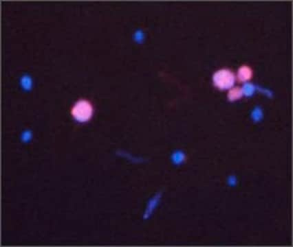
immunofluorescence: suitable
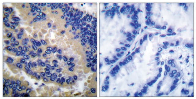
immunohistochemistry (formalin-fixed, paraffin-embedded sections): suitable
western blot: suitable

NCBI accession no.

UniProt accession no.

shipped in

dry ice

target post-translational modification

unmodified

Gene Information

human ... CASP3(836)

General description

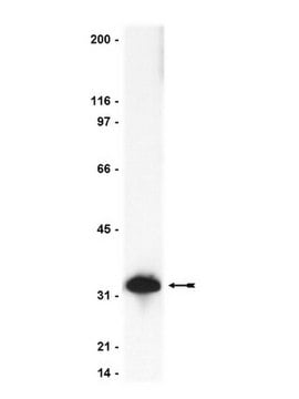
Caspase-3 (CPP32, Apopain) is the most extensively studied apoptotic protein. Caspase-3 is synthesized as an inactive proenzyme (32 kDa) that is processed in cells undergoing apoptosis by self-proteolysis and/or cleavage by another upstream protease. The processed form of caspase-3 consists of large (17 kDa) and small (12 kDa) subunits which associate to form an active enzyme. The active caspase-3 proteolytically cleaves and activates other caspases, as well as relevant targets in the cells such as PARP and DFF.

Specificity

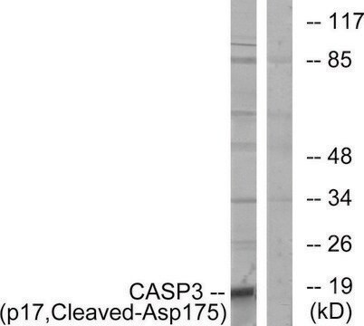
AB3623 detects only the cleaved p17 fragment and does not detect the precursor form in cells undergoing apoptosis.
Reactivity with other species has not been determined.

Immunogen

Cleavage specific synthetic peptide sequence (proprietary).
Epitope: active (cleaved) form

Application

Anti-Caspase 3 Antibody, active (cleaved) form detects level of Caspase 3 and has been published and validated for use in Immunofluorescence (IF), Immunohistochemistry (IHC), and Western Blot (WB).
Research Category
Apoptosis & Cancer

Metabolism
Research Sub Category
Caspases

Enzymes & Biochemistry
Western Blot Analysis:
1:100-1:200 (0.5-4 µg/mL) dilution of active apoptotic cells we used to detect Caspase-3.
Note when using tissue, levels of active caspase 3 can below detectable levels. Use CC119 (human caspase 3) incubated 30 minutes with 1 mM ATP (for activation) as a positive control on westerns. Alternatively, neuronal culture preparations incubated with staurosporine (0.5 µM, 10-18 hr) typically give a strong caspase 3 signal.

Immunohistochemistry:
1:10 dilution (10-20 µg/mL), AB3623 has reacted successfully in 4% PFA fixed, paraffin embedded tissues mouse tissues.

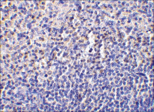
Immunohistochemistry(paraffin):
Optimal Staining of Caspase-3 Monoclonal Antibody: Mouse Small Intestine

Target description

17 kDa

Linkage

Replaces: 04-440; 04-1088

Physical form

ImmunoAffinity Purified
Purified rabbit polyclonal in buffer containing PBS, containing 30% glycerol, 0.5% BSA, 5 mM EDTA and 0.03% proclin.

Storage and Stability

Stable for 1 year at -20ºC from date of receipt.

Analysis Note

Control
Tonsil and appendix tissue. Jurkat cells induced for apoptosis with recombinant soluble TRAIL and staurosporine treated mouse 3T3 cell lysates Skin/Tumor, Embryo.

Other Notes

Concentration: Please refer to the Certificate of Analysis for the lot-specific concentration.

Legal Information

CHEMICON is a registered trademark of Merck KGaA, Darmstadt, Germany

Disclaimer

Unless otherwise stated in our catalog or other company documentation accompanying the product(s), our products are intended for research use only and are not to be used for any other purpose, which includes but is not limited to, unauthorized commercial uses, in vitro diagnostic uses, ex vivo or in vivo therapeutic uses or any type of consumption or application to humans or animals.

Not finding the right product?  

Try our Product Selector Tool.

Pictograms

Exclamation markEnvironment

Signal Word

Warning

Hazard Statements

Hazard Classifications

Aquatic Chronic 2 - Skin Sens. 1

Storage Class Code

10 - Combustible liquids

WGK

WGK 3


Certificates of Analysis (COA)

Search for Certificates of Analysis (COA) by entering the products Lot/Batch Number. Lot and Batch Numbers can be found on a product’s label following the words ‘Lot’ or ‘Batch’.

Already Own This Product?

Find documentation for the products that you have recently purchased in the Document Library.

Visit the Document Library

C Lv et al.
Cell death & disease, 4, e952-e952 (2013-12-07)
Chemotherapy remains the common therapeutic for patients with lung cancer. Novel, selective antitumor agents are pressingly needed. This study is the first to investigate a different, however, effective antitumor drug candidate Wentilactone A (WA) for its development as a novel
Joseph E Druso et al.
The Journal of biological chemistry, 291(17), 8886-8895 (2016-02-26)
The Rho family small GTPase Cdc42 has been implicated in a wide range of cellular functions including the establishment of cell polarity and the remodeling of the actin cytoskeletal architecture, resulting in the tight regulation of cell growth and survival
Hypoxic neuronal necrosis: protein synthesis-independent activation of a cell death program.
Niquet, J; Baldwin, RA; Allen, SG; Fujikawa, DG; Wasterlain, CG
Proceedings of the National Academy of Sciences of the USA null
Martin Breuss et al.
Cell reports, 2(6), 1554-1562 (2012-12-19)
The formation of the mammalian cortex requires the generation, migration, and differentiation of neurons. The vital role that the microtubule cytoskeleton plays in these cellular processes is reflected by the discovery that mutations in various tubulin isotypes cause different neurodevelopmental
Astrocyte proliferation following stroke in the mouse depends on distance from the infarct.
Barreto, GE; Sun, X; Xu, L; Giffard, RG
Testing null

Our team of scientists has experience in all areas of research including Life Science, Material Science, Chemical Synthesis, Chromatography, Analytical and many others.

Contact Technical Service