Skip to Content
Merck
All Photos(3)

Documents

HPA009066

Sigma-Aldrich

Anti-TPM3 antibody produced in rabbit

Prestige Antibodies® Powered by Atlas Antibodies, affinity isolated antibody, buffered aqueous glycerol solution

Synonym(s):

Anti-Tropomyosin α-3 chain antibody produced in rabbit, Anti-Tropomyosin γ antibody produced in rabbit, Anti-Tropomyosin-3 antibody produced in rabbit, Anti-hTM5 antibody produced in rabbit

Sign Into View Organizational & Contract Pricing


About This Item

UNSPSC Code:
12352203
Human Protein Atlas Number:
NACRES:
NA.41

biological source

rabbit

conjugate

unconjugated

antibody form

affinity isolated antibody

antibody product type

primary antibodies

clone

polyclonal

product line

Prestige Antibodies® Powered by Atlas Antibodies

form

buffered aqueous glycerol solution

species reactivity

human, mouse, rat

technique(s)

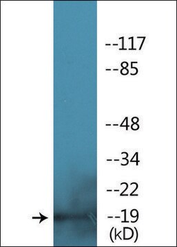
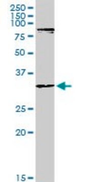
immunoblotting: 0.04-0.4 μg/mL
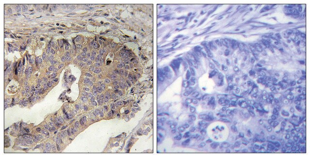
immunohistochemistry: 1:50-1:200

immunogen sequence

AERLQREVEGERRAREQAEAEVASLNRRIQLVEEELDRAQERLATALQKLEEAEKAADESERGMKVIENRALKDEEKMELQEIQLKEAKHIAEEADRKYEEVARKLVIIEGDLERTEERAELAESRCREMDEQIRLMDQNLKCLSA

UniProt accession no.

shipped in

wet ice

storage temp.

−20°C

target post-translational modification

unmodified

Gene Information

human ... TPM3(7170)

General description

TPM3 (tropomyosin 3) gene is localized to human chromosome 1q21.3, and codes for an actin-binding protein. It is present in skeletal and smooth muscles, as well as certain non-muscular tissues. It functions as an oncogene.

Immunogen

Tropomyosin α-3 chain recombinant protein epitope signature tag (PrEST)

Application

Applications in which this antibody has been used successfully, and the associated peer-reviewed papers, are given below.
Western Blotting (1 paper)

Biochem/physiol Actions

TPM3 (tropomyosin 3) is involved in the stabilization of cytoskeleton microfilaments. Upon induction by Ca2+ in skeletal muscles, it mediates the actin-myosin response. However, its function in non-muscular cells is still not clear. TPM3-ALK (anaplastic lymphoma receptor tyrosine kinase) fusion through (1;2) translocation results in hematopoietic tumorigenesis. Fusion with NTRK1 and PDGFRB genes results in the induction of papillary thyroid carcinoma and chronic eosinophilic leukemia. It is responsible for the migration and invasion in hepatocellular carcinoma by facilitating EMT (epithelial-mesenchymal transition) in a Snail-dependent manner. Expression of this protein in tumors is positively linked with the expression of MMP (matrix metalloproteinases). Hence, this protein plays a key role in the progression of gliomas. Mutations in this gene are responsible for the neuromuscular defects in patients with congenital myopathy with fibre type disproportion (CFTD).

Features and Benefits

Prestige Antibodies® are highly characterized and extensively validated antibodies with the added benefit of all available characterization data for each target being accessible via the Human Protein Atlas portal linked just below the product name at the top of this page. The uniqueness and low cross-reactivity of the Prestige Antibodies® to other proteins are due to a thorough selection of antigen regions, affinity purification, and stringent selection. Prestige antigen controls are available for every corresponding Prestige Antibody and can be found in the linkage section.

Every Prestige Antibody is tested in the following ways:
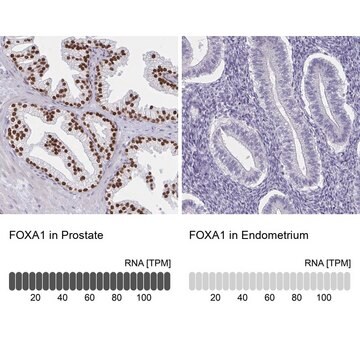
  • IHC tissue array of 44 normal human tissues and 20 of the most common cancer type tissues.
  • Protein array of 364 human recombinant protein fragments.

Linkage

Corresponding Antigen APREST70731

Physical form

Solution in phosphate-buffered saline, pH 7.2, containing 40% glycerol and 0.02% sodium azide

Legal Information

Prestige Antibodies is a registered trademark of Merck KGaA, Darmstadt, Germany

Disclaimer

Unless otherwise stated in our catalog or other company documentation accompanying the product(s), our products are intended for research use only and are not to be used for any other purpose, which includes but is not limited to, unauthorized commercial uses, in vitro diagnostic uses, ex vivo or in vivo therapeutic uses or any type of consumption or application to humans or animals.

Not finding the right product?  

Try our Product Selector Tool.

Storage Class Code

10 - Combustible liquids

WGK

WGK 1

Flash Point(F)

Not applicable

Flash Point(C)

Not applicable

Personal Protective Equipment

dust mask type N95 (US), Eyeshields, Gloves

Certificates of Analysis (COA)

Search for Certificates of Analysis (COA) by entering the products Lot/Batch Number. Lot and Batch Numbers can be found on a product’s label following the words ‘Lot’ or ‘Batch’.

Already Own This Product?

Find documentation for the products that you have recently purchased in the Document Library.

Visit the Document Library

Tao Tao et al.
Tumour biology : the journal of the International Society for Oncodevelopmental Biology and Medicine, 35(9), 9053-9059 (2014-06-11)
Recent studies have shown that many molecular mechanisms, such as the EGFR, AKT, STAT3, and beta-catenin pathways, are involved in glioma. However, the prognosis of the disease remains poor. Explorations of the underlying mechanisms of glioma and identification of effective
P Munot et al.
Neuromuscular disorders : NMD, 20(12), 796-800 (2010-10-19)
Congenital myopathy with fibre type disproportion (CFTD) has been associated with mutations in ACTA1, SEPN1, RYR1 and TPM3 genes. We report the clinico-pathological and electrophysiological features of 2 unrelated cases with heterozygous TPM3 mutation. Case 1 is a 19-year-old lady
Hye-Sun Choi et al.
BMC cancer, 10, 122-122 (2010-04-02)
Since hepatocellular carcinoma (HCC) is one of the leading causes of cancer death worldwide, it is still important to understand hepatocarcinogenesis mechanisms and identify effective markers for tumor progression to improve prognosis. Amplification and overexpression of Tropomyosin3 (TPM3) are frequently
D C Singleton et al.
Oncogene, 34(36), 4713-4722 (2014-12-09)
Hypoxia is a common feature of locally advanced breast cancers that is associated with increased metastasis and poorer survival. Stabilisation of hypoxia-inducible factor-1α (HIF1α) in tumours causes transcriptional changes in numerous genes that function at distinct stages of the metastatic

Our team of scientists has experience in all areas of research including Life Science, Material Science, Chemical Synthesis, Chromatography, Analytical and many others.

Contact Technical Service