Skip to Content
Merck
All Photos(1)

Documents

251M-1

Sigma-Aldrich

Factor XIIIa (AC-1A1) Mouse Monoclonal Antibody

Sign Into View Organizational & Contract Pricing


About This Item

UNSPSC Code:
12352203
NACRES:
NA.41

biological source

mouse

Quality Level

100
500

conjugate

unconjugated

antibody form

diluted ascites fluid

antibody product type

primary antibodies

clone

AC-1A1, monoclonal

description

For In Vitro Diagnostic Use in Select Regions (See Chart)

form

buffered aqueous solution

species reactivity

human

packaging

vial of 0.1 mL concentrate (251M-14)
vial of 0.5 mL concentrate (251M-15)
bottle of 1.0 mL predilute (251M-17)
vial of 1.0 mL concentrate (251M-16)
bottle of 7.0 mL predilute (251M-18)

manufacturer/tradename

Cell Marque

technique(s)

immunohistochemistry (formalin-fixed, paraffin-embedded sections): 1:100-1:500

isotype

IgG1κ

control

dermatofibroma

shipped in

wet ice

storage temp.

2-8°C

visualization

cytoplasmic

Gene Information

human ... F13A1(2162)

General description

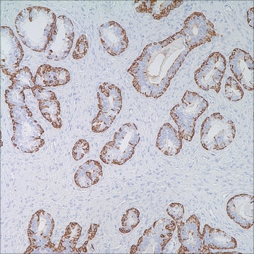
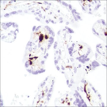
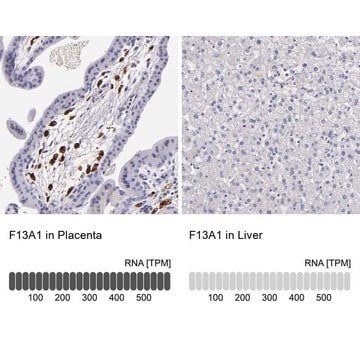
Factor XIIIa is a plasma transglutaminase that is part of the blood coagulation cascade and plays a critical role in stabilizing fibrin networks at the site of an injury to produce a clot. Factor XIIIa is expressed in platelets, megakaryocytes, fibroblast-like cells in the placenta, uterus, and prostate, monocytes and macrophages, and dermal dendritic cells. Anti-Factor XIIIa immunohistochemistry is used to identify fibrohistiocytic neoplasms such as dermatofibroma.

Factor XIIIa is a blood proenzyme that has been identified in platelets, megakaryocyte, and fibroblast-like mesenchymal or histiocytic cells present in the placenta, uterus, and prostate; it is also present in monocytes and macrophages and dermal dendritic cells. Anti- Factor XIIIa has been found to be useful in differentiating between dermatofibroma (90% (+)), dermatofibrosarcoma protuberans (25%(+)), and desmoplastic malignant melanoma (0%(+)). Factor XIIIa positivity is also seen in capillary hemagioblastoma (100%(+)), hemangioendothelioma (100%(+)), hemangiopericytoma (100%(+)), xanthogranuloma (100%(+)), xanthoma (100(+)), hepatocellular carcinoma (93%(+)), glomus tumor (80%(+)), and meningioma (80 % (+)).

Linkage

Factor XIIIa Positive Control Slides , Product No. 251S, are available for immunohistochemistry (formalin-fixed, paraffin-embedded sections).

Physical form

Solution in Tris Buffer, pH 7.3-7.7, with 1% BSA and <0.1% Sodium Azide

Preparation Note

Download the IFU specific to your product lot and formatNote: This requires a keycode which can be found on your packaging or product label.

Other Notes

For Technical Service please contact: 800-665-7284 or email: service@cellmarque.com

Legal Information

Cell Marque is a trademark of Merck KGaA, Darmstadt, Germany

Not finding the right product?  

Try our Product Selector Tool.

Storage Class Code

12 - Non Combustible Liquids

WGK

WGK 2

Flash Point(F)

Not applicable

Flash Point(C)

Not applicable


Certificates of Analysis (COA)

Search for Certificates of Analysis (COA) by entering the products Lot/Batch Number. Lot and Batch Numbers can be found on a product’s label following the words ‘Lot’ or ‘Batch’.

Already Own This Product?

Find documentation for the products that you have recently purchased in the Document Library.

Visit the Document Library

P Abenoza et al.
The American Journal of dermatopathology, 15(5), 429-434 (1993-10-01)
To assess the use of anti-CD34 and anti-factor-XIIIa antibodies for the differential diagnosis of dermatofibroma (DF) and dermatofibrosarcoma protuberans (DFSP), we stained 40 DFs and 13 DFSPs. A significant population of dendritic and spindle cells was reactive with anti-factor-XIIIa in
Z Nemes
Human pathology, 23(7), 805-810 (1992-07-01)
Seven cases of capillary hemangioblastoma from the cerebellum and spinal cord were studied by immunohistochemical methods to determine the origin of the stromal cells. A subpopulation of factor XIIIa-positive tumor cells was a constant feature in hemangioblastomas. These stellate or
E J Glusac et al.
The American journal of surgical pathology, 18(6), 583-590 (1994-06-01)
Epithelioid cell histiocytoma is a recently recognized lesion that is considered to be a variant of cutaneous fibrous histiocytoma (dermatofibroma). Ten cases are presented, including their light microscopic, immunohistochemical, and ultrastructural features. Eight of the cases are similar to those
Louis P Dehner
The American journal of surgical pathology, 27(5), 579-593 (2003-04-30)
Juvenile xanthogranulomas (JXG) is a histiocytic disorder, primarily but not exclusively seen throughout the first two decades of life and principally as a solitary cutaneous lesion. This study is a retrospective clinical and pathologic review of 174 cases documenting the
A J Demetris et al.
The American journal of surgical pathology, 21(3), 263-270 (1997-03-01)
Epithelioid hemangioendothelioma (EHE) is best considered a vascular neoplasm of intermediate malignancy. Although usually progressive, the clinical course is highly unpredictable. The present communication describes a case of extensive recurrent hepatic EHE, limited to the liver allograft and initially manifest

Articles

IHC antibodies enhance dermatopathology beyond H&E stained slides, improving techniques and applications for dermatological research.

Our team of scientists has experience in all areas of research including Life Science, Material Science, Chemical Synthesis, Chromatography, Analytical and many others.

Contact Technical Service