Skip to Content
Merck
All Photos(2)

Documents

ABN194

Sigma-Aldrich

Anti-EVI-5 Antibody

from rabbit, purified by affinity chromatography

Synonym(s):

Ecotropic viral integration site 5 protein homolog, EVI-5

Sign Into View Organizational & Contract Pricing


About This Item

UNSPSC Code:
12352203
eCl@ss:
32160702
NACRES:
NA.41

biological source

rabbit

Quality Level

antibody form

affinity isolated antibody

antibody product type

primary antibodies

clone

polyclonal

purified by

affinity chromatography

species reactivity

mouse, rat

species reactivity (predicted by homology)

human (immunogen homology)

technique(s)

immunohistochemistry: suitable
western blot: suitable

NCBI accession no.

UniProt accession no.

shipped in

wet ice

target post-translational modification

unmodified

Gene Information

human ... EVI5(7813)

General description

Also known as Ecotropic viral integration site 5 protein homolog, EVI-5 serves as a key regulator of the cell cycle through time-dependent stabilization of Emi1, FBX05 protein, and APC/C ubiquitination substrates, such as cyclin A. EVI-5 accumulates in early G1 following Plk1 destruction, but is degraded in a Plk1- and ubiquitin-dependent manner in early mitosis. EVI-5 ablation causes premature APC/C activation, SCF(betaTrCP)-dependent Emi1 degradation, and cyclin destruction, all of which leads to cell cycle arrest and cytokinesis failure. It exists as a monomer and a dimer, and it may form complexes with alpha- and gamma-tubulin or interact with the INCENP-aurora B kinase-survivin chromosomal passenger complex. EVI-5 is implicated in abnormal states, such as multiple sclerosis and neuroblastoma.

Immunogen

KLH-conjugated linear peptide corresponding to the human EVI-5.

Application

Research Category
Neuroscience
Research Sub Category
Signaling Neuroscience
This EVI-5 antibody is validated for use in IHC & WB for the detection of the EVI-5 protein.
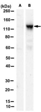
Western Blot Analysis: 2 µg/mL from a representative lot detected EVI-5 in NIH/3T3 nuclear extract.

Quality

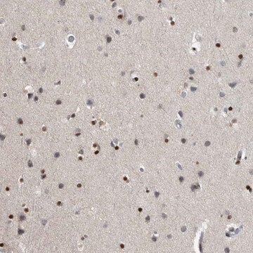
Evaluated by Immunohistochemistry in mouse and rat brain tissues.

Immunohistochemistry Analysis: A 1:500 dilution of this antibody detected EVI-5 in mouse and rat brain tissues.

Target description

~95 kDa observed. Uncharacterized bands may be observed at ~31 kDa, ~17 kDa, and ~15 kDa in some cell lysates. This protein is known to have three potential phosphorylation sites.

Physical form

Affinity purified
Purified rabbit polyclonal in buffer containing 0.1 M Tris-Glycine (pH 7.4), 150 mM NaCl with 0.05% sodium azide.

Storage and Stability

Stable for 1 year at 2-8°C from date of receipt.

Analysis Note

Control
Mouse and rat brain tissues

Other Notes

Concentration: Please refer to the Certificate of Analysis for the lot-specific concentration.

Disclaimer

Unless otherwise stated in our catalog or other company documentation accompanying the product(s), our products are intended for research use only and are not to be used for any other purpose, which includes but is not limited to, unauthorized commercial uses, in vitro diagnostic uses, ex vivo or in vivo therapeutic uses or any type of consumption or application to humans or animals.

Not finding the right product?  

Try our Product Selector Tool.

Storage Class Code

12 - Non Combustible Liquids

WGK

WGK 1

Flash Point(F)

Not applicable

Flash Point(C)

Not applicable


Certificates of Analysis (COA)

Search for Certificates of Analysis (COA) by entering the products Lot/Batch Number. Lot and Batch Numbers can be found on a product’s label following the words ‘Lot’ or ‘Batch’.

Already Own This Product?

Find documentation for the products that you have recently purchased in the Document Library.

Visit the Document Library

Kamila Bedelbaeva et al.
Frontiers in cell and developmental biology, 11, 1101480-1101480 (2023-11-15)
Introduction: The MRL mouse strain is one of the few examples of a mammal capable of healing appendage wounds by regeneration, a process that begins with the formation of a blastema, a structure containing de-differentiating mesenchymal cells. HIF-1α expression in

Our team of scientists has experience in all areas of research including Life Science, Material Science, Chemical Synthesis, Chromatography, Analytical and many others.

Contact Technical Service