Skip to Content
Merck
All Photos(3)

Documents

05-432

Sigma-Aldrich

Anti-NR1 Antibody, CT

Upstate®, from mouse

Synonym(s):

Grin1, NMDA R1 receptor C1 cassette, glutamate receptor, ionotropic, N-methyl D-aspartate 1

Sign Into View Organizational & Contract Pricing


About This Item

UNSPSC Code:
12352203
eCl@ss:
32160702
NACRES:
NA.41

biological source

mouse

Quality Level

antibody form

purified immunoglobulin

antibody product type

primary antibodies

clone

monoclonal

species reactivity

rat

species reactivity (predicted by homology)

human, mouse

manufacturer/tradename

Upstate®

technique(s)

immunocytochemistry: suitable
immunoprecipitation (IP): suitable
western blot: suitable

isotype

IgG

NCBI accession no.

UniProt accession no.

shipped in

wet ice

target post-translational modification

unmodified

Gene Information

human ... GRIN1(2902)
rat ... Grin1(24408)

General description

NR1 (NMDA1or NMDAR1, also known as GRIN1) along with the NR2 subunit forms a heterodimer that make up the NMDA receptor. The NMDA receptor (NMDAR) is characterized as a ionotropic receptor for glutamate. As the name suggests, (NMDA (N-methyl D-aspartate) is an agonist for the receptor. A distinct characteriztic of the NMDA receptor is that it is both ligand-gated and voltage-dependent based on its sensitivity to magnesium. Activated NMDA receptors result in the opening of an ion channel that is nonselective to cations. The result is the flow of Na+ and small amounts of Ca2+ ions into the cell and K+ out of the cell. Calcium flux by the NMDARs has been implicated in synaptic plasticity, a mechanism in the cell for learning and memory.

Specificity

This antibody is specific for NR1, Mr 130 kDa. The epitope maps to a region (834-864) common to all NR1 isoforms and so should cross react with all NR1 isoforms. An additional band may be detected at higher concentrations, Mr 200kDa.

Immunogen

Epitope: The epitope maps to a region (a.a. 834-864) common to all NR1 isoforms
His-tag fusion protein corresponding to residues 834-938 of rat NR1A. The immunizing sequence is identical in rat and human.

Application

Anti-NR1 Antibody, CT detects level of NR1 & has been published & validated for use in IC, IP & WB.
Immunoprecipitation:
4 μg of a previous lot immunoprecipitated NR1 from 500mg of rat brain microsomal protein preparation.

Immunocytochemistry:
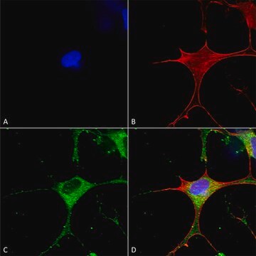
An independent laboratory has shown positive staining in QT-6 cells, transfected to express NR1, which were fixed with 4% paraformaldehyde/4% sucrose in PBS and in nontransfected cultured rat neurons.

Note: Do not boil the microsomal preparation. Incubate at room temperature for 30-45 minutes.

Quality

Routinely evaluated by Western Blot on rat brain microsomal protein preparation.

Western Blot Analysis:
0.5-2 μg/mL of this lot detected NR1 from 20 μg of rat brain microsomal protein preparation (Catalog # 12-144).

Target description

130 kDa observed

Physical form

Format: Purified
Purified mouse monoclonal IgG in buffer containing 0.1M Tris-glycine, pH 7.4, 0.15M NaCl, 0.05% sodium azide.

Storage and Stability

Stable for 1 year at 2° to 8ºC from date of receipt.

Other Notes

Concentration: Please refer to the Certificate of Analysis for the lot-specific concentration.

Legal Information

UPSTATE is a registered trademark of Merck KGaA, Darmstadt, Germany

Not finding the right product?  

Try our Product Selector Tool.

Storage Class Code

10 - Combustible liquids

WGK

WGK 1


Certificates of Analysis (COA)

Search for Certificates of Analysis (COA) by entering the products Lot/Batch Number. Lot and Batch Numbers can be found on a product’s label following the words ‘Lot’ or ‘Batch’.

Already Own This Product?

Find documentation for the products that you have recently purchased in the Document Library.

Visit the Document Library

Glutamate binding to the GluN2B subunit controls surface trafficking of N-methyl-D-aspartate (NMDA) receptors.
She, K; Ferreira, JS; Carvalho, AL; Craig, AM
The Journal of Biological Chemistry null
Natural reward experience alters AMPA and NMDA receptor distribution and function in the nucleus accumbens.
Pitchers, KK; Schmid, S; Di Sebastiano, AR; Wang, X; Laviolette, SR; Lehman, MN; Coolen, LM
Testing null
Jia-Hua Hu et al.
Nature neuroscience, 15(6), 836-844 (2012-05-09)
Group I metabotropic glutamate receptors (mGluRs), including mGluR1 and mGluR5, are G protein–coupled receptors (GPCRs) that are expressed at excitatory synapses in brain and spinal cord. GPCRs are often negatively regulated by specific G protein–coupled receptor kinases and subsequent binding
NMDA receptors mediate synaptic competition in culture.
She, K; Craig, AM
Testing null
Kourtney Graham et al.
NPJ schizophrenia, 5(1), 1-1 (2019-01-16)
The 14-3-3 family of proteins is genetically linked to several psychiatric disorders, including schizophrenia. Our 14-3-3 functional knockout (FKO) mice, as well as other 14-3-3 knockout models, have been shown to exhibit behavioral endophenotypes related to schizophrenia. While specific forebrain

Our team of scientists has experience in all areas of research including Life Science, Material Science, Chemical Synthesis, Chromatography, Analytical and many others.

Contact Technical Service