Skip to Content
Merck
All Photos(3)

Documents

MAB079-1

Sigma-Aldrich

Anti-S-100 Protein Antibody, clone 15E2E2

clone 15E2E2, Chemicon®, from mouse

Synonym(s):

Calcium Binding Protein A1/B1

Sign Into View Organizational & Contract Pricing


About This Item

UNSPSC Code:
12352203
eCl@ss:
32160702
NACRES:
NA.41

biological source

mouse

Quality Level

antibody form

purified immunoglobulin

antibody product type

primary antibodies

clone

15E2E2, monoclonal

species reactivity

bovine, rat, human, mouse

manufacturer/tradename

Chemicon®

technique(s)

immunohistochemistry (formalin-fixed, paraffin-embedded sections): suitable
immunoprecipitation (IP): suitable
western blot: suitable

isotype

IgG2aκ

NCBI accession no.

UniProt accession no.

shipped in

wet ice

target post-translational modification

unmodified

Gene Information

human ... S100A1(6271)

General description

S-100 protein is a member of the EF-hand calcium-binding protein superfamily that is widely distributed and conserved in the central nervous system of vertebrates. The protein exists in both heterodimeric and homodimeric forms. The two subunits of S-100 protein are the products of separate genes that are differentially expressed by various cells. The beta subunit is present in all S-100 positive cells and tumors. In contrast, the alpha subunit is detectable only in heart, skeletal muscle and brain. In addition to its calcium binding properties, S-100 protein has a high-affinity binding site for zinc and is involved in the regulation of protein phosphorylation in the brain. The protein apparently plays a role in cell differentiation and growth, cytoskeletal structure and function, and has been implicated in neuropathological diseases.

Specificity

This antibody recognizes ~22 kDa (non-reduced) or 11 kDa (reduced) of S100. It reacts with human, rat, mouse, and cow (bovine). Other species are not tested.

Immunogen

Epitope: Recognizes both alpha & beta S100
Purified bovine S-100 protein

Application

Anti-S-100 Protein Antibody, clone 15E2E2 is an antibody against S-100 Protein for use in IH, IH(P), IP & WB.
Immunoprecipitation:
A previous lot of this antibody was used in IP.

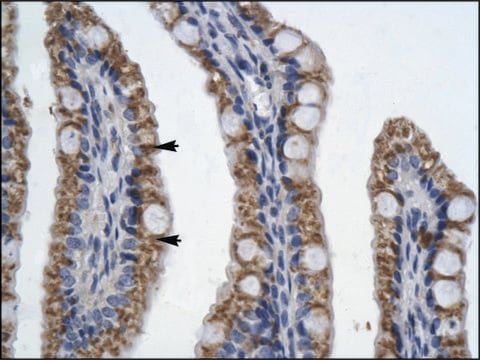
Visualization of cellular and subcellular localization of S-100 protein using immunohistochemical techniques.

Immunohistochemistry:
A 1:100-200 dilution of a previous lot was used for paraffin embedded tissue sections.

Optimal working dilutions must be determined by end user.
Research Category
Metabolism
Research Sub Category
Muscle Physiology

Quality

Evaluated by Western Blot on PC12 lysates

Western Blot Analysis:
1:1000 dilution of this antibody detected S100 on 10 µg of PC12 lysate.

Physical form

Format: Purified
The mouse IgG is purified by protein A-affinity chromatography according to isotyping.
This affinity purified antibody is supplied in sterile phosphate buffered saline (pH 7.2) containing antibody stabilizer. Contains 0.01% sodium azide.

Storage and Stability

Stable for 1 year at 2-8ºC from date of receipt.

Analysis Note

Control
Positive Control Tissue: Melanoma

Other Notes

Concentration: 0.5 mg/mL

Legal Information

CHEMICON is a registered trademark of Merck KGaA, Darmstadt, Germany

Disclaimer

Unless otherwise stated in our catalog or other company documentation accompanying the product(s), our products are intended for research use only and are not to be used for any other purpose, which includes but is not limited to, unauthorized commercial uses, in vitro diagnostic uses, ex vivo or in vivo therapeutic uses or any type of consumption or application to humans or animals.

Not finding the right product?  

Try our Product Selector Tool.

Storage Class Code

12 - Non Combustible Liquids

WGK

WGK 2

Flash Point(F)

Not applicable

Flash Point(C)

Not applicable


Certificates of Analysis (COA)

Search for Certificates of Analysis (COA) by entering the products Lot/Batch Number. Lot and Batch Numbers can be found on a product’s label following the words ‘Lot’ or ‘Batch’.

Already Own This Product?

Find documentation for the products that you have recently purchased in the Document Library.

Visit the Document Library

Jonathan D Verrier et al.
The Journal of pharmacology and experimental therapeutics, 354(2), 175-183 (2015-05-23)
The 3',5'-cAMP-adenosine pathway (3',5'-cAMP→5'-AMP→adenosine) and the 2',3'-cAMP-adenosine pathway (2',3'-cAMP→2'-AMP/3'-AMP→adenosine) are active in the brain. Oligodendrocytes participate in the brain 2',3'-cAMP-adenosine pathway via their robust expression of 2',3'-cyclic nucleotide 3'-phosphodiesterase (CNPase; converts 2',3'-cAMP to 2'-AMP). Because Schwann cells also express CNPase
Steven Petratos et al.
Journal of neuropathology and experimental neurology, 62(4), 398-411 (2003-05-02)
Schwann cell death is a developmentally regulated phenomenon and is also induced after peripheral nerve axotomy in neonatal rodents. In this study, we explored whether ligand-induced activation of the low-affinity neurotrophin receptor (p75(NTR)) is responsible for inducing Schwann cell death
Giant schwannoma of the cheek--a comprehensive histological and immunohistochemical description of a rare tumour.
Mariusz Gajda, Olaf Zagolski, Agnieszka Jasztal, Grzegorz J Lis, Dariusz Adamek et al.
Polish Journal of Pathology : Official Journal of the Polish Society of Pathologists null
Immunohistochemical visualization of neurons and specific glial cells for stereological application in the porcine neocortex.
Lise Lyck, Jacob Jelsing, Pia S?ndergaard Jensen, Kate Lykke Lambertsen, Bente Pakkenberg et al.
Journal of Neuroscience Methods null
Cellular immunolocalization of S100 protein within fixed tissue sections by monoclonal antibodies.
Loeffel, S C, et al.
Archives of Pathology & Laboratory Medicine, 109, 117-122 (1985)

Our team of scientists has experience in all areas of research including Life Science, Material Science, Chemical Synthesis, Chromatography, Analytical and many others.

Contact Technical Service