Skip to Content
Merck
All Photos(4)

Documents

F3777

Sigma-Aldrich

Anti-Actin, α-Smooth Muscle - FITC antibody, Mouse monoclonal

clone 1A4, purified from hybridoma cell culture

Synonym(s):

Monoclonal Anti-Actin, α-Smooth Muscle, alpha SMA-FITC antibody, alpha smooth muscle actin - FITC antibody, SMA

Sign Into View Organizational & Contract Pricing


About This Item

UNSPSC Code:
12352203
NACRES:
NA.41

biological source

mouse

conjugate

FITC conjugate

antibody form

purified from hybridoma cell culture

antibody product type

primary antibodies

clone

1A4, monoclonal

form

buffered aqueous solution

mol wt

antigen ~42 kDa

species reactivity

human, mouse, rat, chicken, frog, canine, rabbit, guinea pig, goat, bovine, sheep, snake

technique(s)

immunohistochemistry (formalin-fixed, paraffin-embedded sections): 1:500 using human tonsil or appendix

isotype

IgG2a

UniProt accession no.

shipped in

dry ice

storage temp.

−20°C

target post-translational modification

unmodified

Gene Information

human ... ACTA2(59)
mouse ... Acta2(11475)
rat ... Acta2(81633)

Looking for similar products? Visit Product Comparison Guide

General description

α-smooth muscle actin, a smooth muscle cell-specific protein, is also known as aortic smooth muscle or a smooth muscle actin (α-SMA, SMactin, α-SM-actin, ASMA). It belongs to the actin family.It is predominantly found in cells of the smooth muscle lineages. It has been reported that α-smooth muscle actin was also found in certain non-muscle cells, in particular fibroblasts i.e. myofibroblasts. Actin, a conserved eukaryotic protein, is the major cytoskeletal component and is expressed as six different isoforms in mammals and birds. It is mapped to human chromosome 10q23.31.

The antibody reacts with normal and neoplastic, human vascular and visceral smooth muscle cells. It reacts with normal myoepithelial cells, pericytes, eye lens cells, hair follicle cells and certain stromal cells in the intestine, testis, lymphoid organs, liver, ovary and bone marrow. The antibody also reacts with stromal myofibroblasts in hypertrophic scars, and in neoplastic tissues. α-smooth muscle actin is transiently co-expressed with sarcomeric α-actin during myogenesis in chicken and rat embryos. It was also found in the ventricular conducting tract of adult mammalian heart.
The antibody (also known as anti-α-Sm-1) is specific for the single isoform of α-smooth muscle actin. It reacts specifically with α-smooth muscle actin in immunoblotting assays and labels smooth muscle cells in frozen or formalin-fixed, paraffin-embedded tissue sections.

Specificity

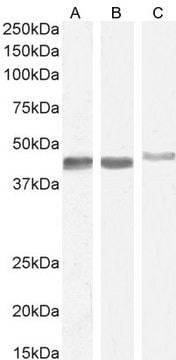
Monoclonal Anti-Actin, α-Smooth Muscle specifically recognizes the α-smooth muscle isoform of actin (42 kDa) by ELISA and immunoblotting. It does not react with the other major actin isoforms present in fibroblasts or epithelial cells (β and γ-cytoplasmic), striated muscle (α-sarcomeric), myocardium (α-myocardial), or γ-smooth muscle isoform.

Immunogen

N-terminal synthetic decapeptide of α-smooth muscle actin.

Application

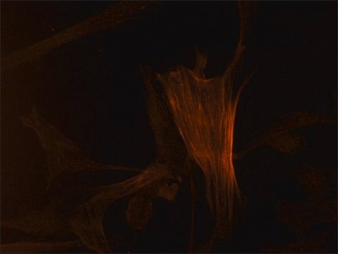
Immunocytochemistry was performed on smooth muscle cells from bovine aortas using the monoclonal anti-ACTA2 antibody. Cells were first grown on glass cover slips and fixed in 50% acetone/EtOH for 10 minutes at 4 degrees.
Monoclonal Anti-Actin, α-Smooth Muscle - FITC antibody is suitable for:
  • immunohistochemical (IHC) staining of heart, lung, kidney, and liver of mice to examine the PHD2 deficiency in internal organs
  • immunostaining to analyze vascular smooth muscle cells/pericytes development in platelet-derived growth factor (PDGFR) and PDGFR- β-/- embryos
  • immunofluorescent analysis of implanted cells, frozen tissue samples to study the similar levels of expression of SMaA and SM-MHC as established primary SMC lines
  • western blots of lens epithelial cells to determine the distribution of α-SMA expression in lens epithelial cell
  • Immunohistochemical examination of lesion morphology
  • flow cytometry
  • immunohistochemistry, proliferation and TUNEL (terminal deoxynucleotidyl transferase (TdT) dUTP nick-end labeling) assays
  • immunocytochemistry
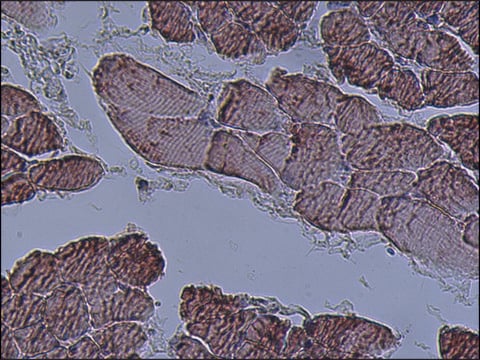
Paraffin embedded sections of rat testis tissue grafts were immunohistochemically stained with mouse monoclonal anti-smooth muscle actin.
IHC analysis of x-gal stained muouse cardiac tissue was performed using the primary antibody, mouse monoclonal anti-smooth muscle actin to identify myofibroblasts.

Biochem/physiol Actions

Actin plays an important role in cell motility, structure and integrity. Smooth muscle actin-α (SMA)/ α2-smooth muscle actin (ACTA2) interacts with β-myosin heavy chain and helps in vascular smooth muscle cell contraction. The encoded protein modulates the expression of c-MET (tyrosine-protein kinase Met) and focal adhesion kinase (FAK) in human lung adenocarcinoma cells, which positively and selectively mediates tumor progression. Mutation in the gene expression leads to thoracic aortic aneurysms and dissections (TAAD).

Physical form

Solution in 0.01 M phosphate buffered saline, pH 7.4, containing 1% bovine serum albumin and 15 mM sodium azide, as a preservative.

Storage and Stability

For continuous use, store at 2-8 °C for up to one month. For extended storage, freeze in working aliquots. Repeated freezing and thawing, or storage in "frostfree" freezers, is not recommended. If slight turbidity occurs upon prolonged storage, clarify the solution by centrifugation before use. Protect from prolonged exposure to light.

Other Notes

To view an Actin antibody selection guide, please visit www.sigmaaldrich.com/actin.

Disclaimer

Unless otherwise stated in our catalog or other company documentation accompanying the product(s), our products are intended for research use only and are not to be used for any other purpose, which includes but is not limited to, unauthorized commercial uses, in vitro diagnostic uses, ex vivo or in vivo therapeutic uses or any type of consumption or application to humans or animals.

Not finding the right product?  

Try our Product Selector Tool.

Storage Class Code

12 - Non Combustible Liquids

WGK

nwg

Flash Point(F)

Not applicable

Flash Point(C)

Not applicable

Personal Protective Equipment

dust mask type N95 (US), Eyeshields, Gloves

Certificates of Analysis (COA)

Search for Certificates of Analysis (COA) by entering the products Lot/Batch Number. Lot and Batch Numbers can be found on a product’s label following the words ‘Lot’ or ‘Batch’.

Already Own This Product?

Find documentation for the products that you have recently purchased in the Document Library.

Visit the Document Library

Modification of proteins by norepinephrine is important for vascular contraction.
Johnson K B, et al.
Frontiers in Physiology, 1, 131-131 (2010)
Tbx18 function in epicardial development.
Greulich F, et al.
Cardiovascular Research, 96(3), 476-483 (2012)
AICAR-dependent AMPK activation improves scar formation in the aged heart in a murine model of reperfused myocardial infarction.
Cieslik K A, et al.
Journal of Molecular and Cellular Cardiology, 63, 26-36 (2013)
Jiusong Sun et al.
Circulation, 120(11), 973-982 (2009-09-02)
Mast cell chymase may participate in the pathogenesis of human abdominal aortic aneurysm (AAA), yet a direct contribution of this serine protease to AAA formation remains unknown. Human AAA lesions had high numbers of chymase-immunoreactive mast cells. Serum chymase level
Kai Schuh et al.
The Journal of biological chemistry, 278(42), 41246-41252 (2003-08-06)
The mechanisms governing vascular smooth muscle tone are incompletely understood. In particular, the role of the sarcolemmal calcium pump PMCA (plasma membrane calmodulin-dependent calcium ATPase), which extrudes Ca2+ from the cytosol, and its importance compared with the sodium/calcium exchanger remain

Our team of scientists has experience in all areas of research including Life Science, Material Science, Chemical Synthesis, Chromatography, Analytical and many others.

Contact Technical Service