05-806
Anti-phospho-Histone H3 (Ser10) Antibody, clone 3H10
clone 3H10, Upstate®, from mouse
Synonym(s):
H3 histone, family 3A, H3S10P, Histone H3 (phospho S10), H3 histone, family 3A, H3 histone, family 3B, H3 histone, family 3B (H3.3B)
About This Item
Recommended Products
biological source
mouse
Quality Level
antibody form
purified immunoglobulin
antibody product type
primary antibodies
clone
3H10, monoclonal
species reactivity
human
manufacturer/tradename
Upstate®
technique(s)
flow cytometry: suitable
immunocytochemistry: suitable
immunofluorescence: suitable
inhibition assay: suitable (peptide)
multiplexing: suitable
western blot: suitable
isotype
IgG1κ
NCBI accession no.
UniProt accession no.
shipped in
wet ice
target post-translational modification
phosphorylation (pSer10)
Gene Information
human ... HIST1H3F(8968)
General description
Specificity
Application
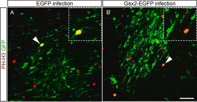
This antibody has been reported by an independent laboratory to detect phosphorylated histone H3 using flow cytometry.
Beadlyte Histone-Peptide Specificity Assay:
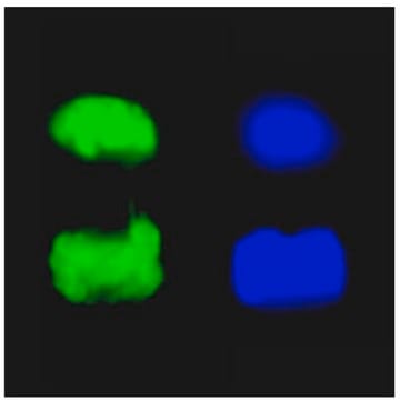
1:1,000 to 1:81,000 dilutions of a previous lot were incubated with histone H3 peptides containing various modifications conjugated to Luminex microspheres. (Figure B). Only the peptide containing phospho-serine 10 was detected.
Immunocytochemistry:
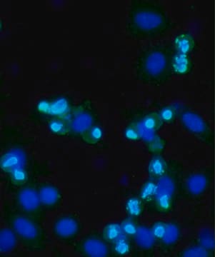
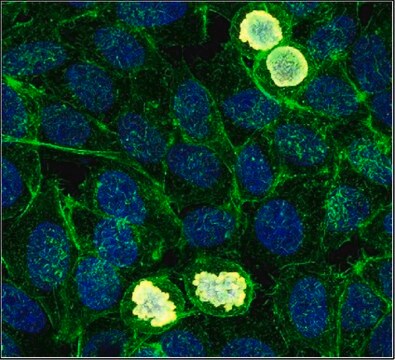
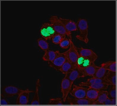
0.2 μg/mL of a previous lot showed positive chromosome immunostaining for mitotic A431 and HeLa cells fixed with 95% ethanol and 5% acetic acid and permeabilized with 0.1% Triton X-100.
Peptide Inhibition Analysis:
Detection of histone H3 in immunoblots of colcemid treated HeLa acid extracts by anti-phospho-Histone H3 (Ser10) was diminished by 10 μM of histone H3 peptide containing phospho-serine 10, but not by peptides containing phospho-serine 28 or an unmodified histone H3 sequence (Figure C).
Immunofluorescence:
Quality
Western Blot Analysis:
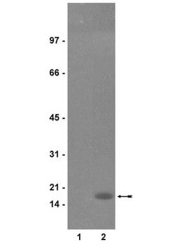
0.05-0.5 μg/mL of this lot detected phosphorylated histone H3 in acid extracted proteins from mitotic HeLa cells treated with colcemid (Catalog # 17-306), but not unmodified recombinant Histone H3 (Catalog # 14-494) (Figure A).
Target description
Physical form
Storage and Stability
Handling Recommendations:
Upon receipt, and prior to removing the cap, centrifuge the vial and gently mix the solution. Aliquot into microcentrifuge tubes and store at -20°C. Avoid repeated freeze/thaw cycles, which may damage IgG and affect product performance. Note: Variability in freezer temperatures below -20°C may cause glycerol containing solutions to become frozen during storage.
Analysis Note
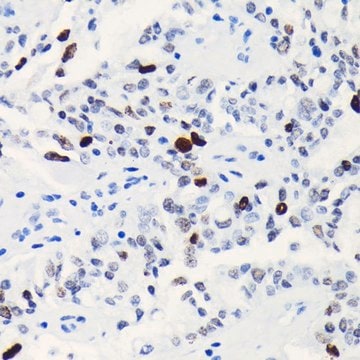
UV-treated 293 cell extracts, UV-treated HeLa cell extracts, breast cancer tissue, HEPG2 cell extracts.
Other Notes
Legal Information
Not finding the right product?
Try our Product Selector Tool.
recommended
Storage Class Code
12 - Non Combustible Liquids
WGK
WGK 1
Flash Point(F)
Not applicable
Flash Point(C)
Not applicable
Certificates of Analysis (COA)
Search for Certificates of Analysis (COA) by entering the products Lot/Batch Number. Lot and Batch Numbers can be found on a product’s label following the words ‘Lot’ or ‘Batch’.
Already Own This Product?
Find documentation for the products that you have recently purchased in the Document Library.
Our team of scientists has experience in all areas of research including Life Science, Material Science, Chemical Synthesis, Chromatography, Analytical and many others.
Contact Technical Service