Skip to Content
Merck
All Photos(1)

Key Documents

MABE1123

Sigma-Aldrich

Anti-XPB Antibody, clone 15TF2-1B3

ascites fluid, clone 15TF2-1B3, from mouse

Synonym(s):

TFIIH basal transcription factor complex helicase XPB subunit, Basic transcription factor 2 89 kDa subunit, BTF2 p89, DNA excision repair protein ERCC-3, DNA repair protein complementing XP-B cells, TFIIH basal transcription factor complex 89 kDa subunit

Sign Into View Organizational & Contract Pricing


About This Item

UNSPSC Code:
12352203
eCl@ss:
32160702
NACRES:
NA.41
clone:
15TF2-1B3, monoclonal
application:
ICC
WB
species reactivity:
human
technique(s):
immunocytochemistry: suitable
western blot: suitable
citations:
4

biological source

mouse

Quality Level

antibody form

ascites fluid

antibody product type

primary antibodies

clone

15TF2-1B3, monoclonal

species reactivity

human

technique(s)

immunocytochemistry: suitable
western blot: suitable

isotype

IgG1κ

NCBI accession no.

UniProt accession no.

shipped in

wet ice

target post-translational modification

unmodified

Gene Information

human ... ERCC3(2071)

General description

Transcription factor II human (TFIIH) basal transcription factor complex helicase XPB subunit (EC 3.6.4.12; UniProt P19447; also known as BTF2 p89, DNA excision repair protein ERCC-3, DNA repair helicase, DNA repair protein complementing XP-B cells, TFIIH 89 kDa subunit, TFIIH basal transcription factor complex 89 kDa subunit, TFIIH p89, Basic transcription factor 2 89 kDa subunit, Xeroderma pigmentosum group B-complementing protein) is encoded by the ERCC3 (also known as BTF2, GTF2H, RAD25, TFIIH, XPB) gene (Gene ID 2071) in human. DNA lesions caused by UV irradiation, drugs, or other environmental factors are eliminated by two nucleotide excision repair (NER) pathways, Global genome repair (GGR) and transcription-coupled repair (TCR). In GGR, the removal of lesions requires their recognition by the repair factor XPC/HR23b and the subsequent opening of the DNA duplex by TFIIH. The resulting single-stranded structure is stabilized by XPA and replication protein A (RPA). XPG is recruited through its interaction with TFIIH on the 3′ side of the lesion and its positioning on the cut site requires RPA. The interaction between XPA and XPB (ERCC1) stimulates the recruitment of ERCC1-XPF on the 5′ side of the DNA lesion. The damaged oligonucleotide can then be removed through the double incision by XPG and ERCC1-XPF endonucleases. In TCR, these factors (except XPC/HR23B) are recruited by the stalled RNA pol II in front of the damage with the help of the CSB and CSA proteins.

Immunogen

Epitope: N-terminus
Recombinant protein corresponding to human XPB.

Application

Research Category
Epigenetics & Nuclear Function
Research Sub Category
Nuclear Receptors
This Anti-XPB Antibody, clone 15TF2-1B3 is validated for use in Western Blotting, Immunocytochemistry for the detection of XPB.
Western Blotting Analysis: A representative lot detected Xpb in murine embryonic fibroblasts (MEFs) and HeLa cells, as well as in transgenic animal-derived MEFs expressing Xpb lacking last 43 C-terminal amino acids (Andressoo, J.O., et al. (2009). Mol Cell Biol.29(5):1276-290).
Western Blotting Analysis: A representative lot detected endougenous as well as exogenously expressed Xpb in both U2OS17 whole cell lysate and in THIIF p62 subunit immunoprecipitate (Ziani, S., et al. (2014). J Cell Biol.;206(5):589-598).
Western Blotting Analysis: A representative lot detected Xpb in THIIF TTDA subunit immunoprecipitate (Giglia-Mari,G., et al. (2006). PLoS Biol. 4(6): e156).
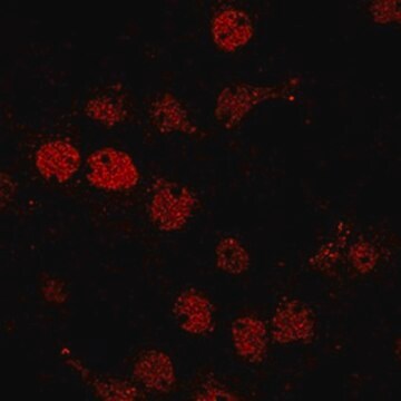
Immunocytochemistry Analysis: A representative lot detected XPB recruitment to the DNA damage sites in the nuclei of UV-irradated HeLa cells (Alekseev, S., et al. (2014). Chem Biol. 21(3):398-407).

Quality

Evaluated by Western Blotting in HeLa nuclear extract.

Western Blotting Analysis: A 1:2,000 dilution of this antibody detected XPB in 10 µg of HeLa nuclear extract.

Target description

~85 kDa observed. Uncharacterized band(s) may appear in some lysates.

Physical form

Mouse monoclonal IgG1κ ascites with 0.05% sodium azide.
Unpurified

Storage and Stability

Stable for 1 year at -20°C from date of receipt.
Handling Recommendations: Upon receipt and prior to removing the cap, centrifuge the vial and gently mix the solution. Aliquot into microcentrifuge tubes and store at -20°C. Avoid repeated freeze/thaw cycles, which may damage IgG and affect product performance.

Other Notes

Concentration: Please refer to lot specific datasheet.

Disclaimer

Unless otherwise stated in our catalog or other company documentation accompanying the product(s), our products are intended for research use only and are not to be used for any other purpose, which includes but is not limited to, unauthorized commercial uses, in vitro diagnostic uses, ex vivo or in vivo therapeutic uses or any type of consumption or application to humans or animals.

Not finding the right product?  

Try our Product Selector Tool.

Storage Class Code

12 - Non Combustible Liquids

WGK

nwg

Flash Point(F)

Not applicable

Flash Point(C)

Not applicable


Certificates of Analysis (COA)

Search for Certificates of Analysis (COA) by entering the products Lot/Batch Number. Lot and Batch Numbers can be found on a product’s label following the words ‘Lot’ or ‘Batch’.

Already Own This Product?

Find documentation for the products that you have recently purchased in the Document Library.

Visit the Document Library

Sylvain Geny et al.
Methods in molecular biology (Clifton, N.J.), 2247, 39-57 (2020-12-11)
Macromolecular complexes govern the majority of biological processes and are of great biomedical relevance as factors that perturb interaction networks underlie a number of diseases, and inhibition of protein-protein interactions is a common strategy in drug discovery. Genome editing technologies
Julian Haase et al.
eLife, 11 (2022-03-17)
Condensins compact chromosomes to promote their equal segregation during mitosis, but the mechanism of condensin engagement with and action on chromatin is incompletely understood. Here, we show that the general transcription factor TFIIH complex is continuously required to establish and
Masanobu Ueda et al.
Genes to cells : devoted to molecular & cellular mechanisms, 24(4), 284-296 (2019-02-15)
The multisubunit complex transcription factor IIH (TFIIH) has dual functions in transcriptional initiation and nucleotide excision repair (NER). TFIIH is comprised of two subcomplexes, the core subcomplex (seven subunits) including XPB and XPD helicases and the cyclin-dependent kinase (CDK)-activating kinase
Dinesh Verma et al.
Proceedings of the National Academy of Sciences of the United States of America, 117(23), 13044-13055 (2020-05-22)
Epstein-Barr virus (EBV) is associated with epithelial and lymphoid malignancies, establishes latent infection in memory B cells, and intermittently produces infectious virions through lytic replication. Released virions play a key role in latent reservoir maintenance and transmission. Lytic EBV transcription

Our team of scientists has experience in all areas of research including Life Science, Material Science, Chemical Synthesis, Chromatography, Analytical and many others.

Contact Technical Service