Skip to Content
Merck
All Photos(3)

Key Documents

SAB4200130

Sigma-Aldrich

Anti-Phospholipase A2 (iPLA2) (C-terminal region) antibody produced in rabbit

enhanced validation

~1.5 mg/mL, affinity isolated antibody

Synonym(s):

Anti-CaI-PLA2, Anti-INAD1, Anti-IPLA2-VIA, Anti-PARK14, Anti-PLA2, Anti-PLA2G6, Anti-PNPLA9, Anti-Phospholipase A2, group VI (cytosolic, calcium-independent), GVI

Sign Into View Organizational & Contract Pricing


About This Item

MDL number:
UNSPSC Code:
12352203
NACRES:
NA.41
conjugate:
unconjugated
application:
WB
clone:
polyclonal
species reactivity:
mouse, human, rat
citations:
7
technique(s):
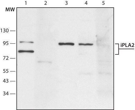
western blot: 1-2 μg/mL using extract of HEK-293T cells overexpressing human iPLA2, and 1-2 mg/mL using rat kidney extract (S1 fraction).
western blot: 1-2 μg/mL using rat kidney extract (S1 fraction)

biological source

rabbit

conjugate

unconjugated

antibody form

affinity isolated antibody

antibody product type

primary antibodies

clone

polyclonal

form

buffered aqueous solution

mol wt

antigen ~85 kDa
antigen ~95 kDa

species reactivity

mouse, human, rat

packaging

antibody small pack of 25 μL

enhanced validation

recombinant expression
Learn more about Antibody Enhanced Validation

concentration

~1.5 mg/mL

technique(s)

western blot: 1-2 μg/mL using extract of HEK-293T cells overexpressing human iPLA2, and 1-2 mg/mL using rat kidney extract (S1 fraction).
western blot: 1-2 μg/mL using rat kidney extract (S1 fraction)

UniProt accession no.

shipped in

dry ice

storage temp.

−20°C

target post-translational modification

unmodified

Gene Information

human ... PLA2G6(8398)

General description

Ca2+-independent phospholipase A2 (iPLA2), also known as PLA2G6, is a member of the PLA2 superfamily. It consists of a C-terminal calmodulin-binding domain, a bipartite nuclear localization sequence, GXSXG serine lipase residues, and ankyrin repeats at the N-terminus. iPLA2 gene is mapped to human chromosome 22q13.1 and encodes alternatively spliced isoforms.

Specificity

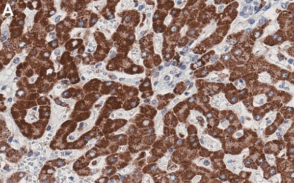
Anti-Phospholipase A2 (iPLA2) (C-terminal region), specifically recognizes human and rat iPLA2.

Application

Anti-Phospholipase A2 (iPLA2) (C-terminal region) antibody produced in rabbit has been used in immunoblotting.

Biochem/physiol Actions

Ca2+-independent phospholipase A2 (iPLA2) catalyzes the cleavage of fatty acids from the sn-2 position of phospholipids. iPLA2 is implicated in leukotriene and prostaglandin production as well as in arachidonic acid release phospholipid remodeling. Mutations in the PLA2G6 gene are the cause of two childhood neurologic disorders, neurodegeneration with brain iron accumulation (NBIA) and infantile neuroaxonal dystrophy 1 (INAD1). Recent evidence suggests that both Ca2+-dependent PLA2 (cPLA2) and iPLA2 may play a central role in memory deficits at early stages.

Physical form

Solution in 0.01 M phos­phate buffered saline, pH 7.4, containing 15 mM sodium azide.

Storage and Stability

Store at −20 °C. For continuous use, the product may be stored at 2−8 °C for up to one month. For extended storage, freeze at −20 °C in working aliquots. Repeated freezing and thawing, or storage in “frost-free” freezers, is not recommended. If slight turbidity occurs upon prolonged storage, clarify the solution by centrifugation. Discard working dilutions if not used within 12 hours.

Disclaimer

Unless otherwise stated in our catalog or other company documentation accompanying the product(s), our products are intended for research use only and are not to be used for any other purpose, which includes but is not limited to, unauthorized commercial uses, in vitro diagnostic uses, ex vivo or in vivo therapeutic uses or any type of consumption or application to humans or animals.

Not finding the right product?  

Try our Product Selector Tool.

Storage Class Code

10 - Combustible liquids

Flash Point(F)

Not applicable

Flash Point(C)

Not applicable


Choose from one of the most recent versions:

Certificates of Analysis (COA)

Lot/Batch Number

Don't see the Right Version?

If you require a particular version, you can look up a specific certificate by the Lot or Batch number.

Already Own This Product?

Find documentation for the products that you have recently purchased in the Document Library.

Visit the Document Library

Laura A Engel et al.
PloS one, 5(9), e12897-e12897 (2010-10-05)
Mutations in the PLA2G6 gene have been identified in autosomal recessive neurodegenerative diseases classified as infantile neuroaxonal dystrophy (INAD), neurodegeneration with brain iron accumulation (NBIA), and dystonia-parkinsonism. These clinical syndromes display two significantly different disease phenotypes. NBIA and INAD are
Zhengshan Zhao et al.
PloS one, 6(10), e26991-e26991 (2011-11-03)
Infantile neuroaxonal dystrophy (INAD) is a progressive, autosomal recessive neurodegenerative disease characterized by axonal dystrophy, abnormal iron deposition and cerebellar atrophy. This disease was recently mapped to PLA2G6, which encodes group VI Ca(2+)-independent phospholipase A(2) (iPLA(2) or iPLA(2)β). Here we
Evelin L Schaeffer et al.
Psychopharmacology, 198(1), 1-27 (2008-04-09)
Alzheimer disease (AD), a progressive neurodegenerative disorder, is the leading cause of dementia in the elderly. A combination of cholinergic and glutamatergic dysfunction appears to underlie the symptomatology of AD, and thus, treatment strategies should address impairments in both systems.
Govind Gupta et al.
ACS nano, 17(17), 17451-17467 (2023-08-29)
Nanoparticles (NPs) elicit sterile inflammation, but the underlying signaling pathways are poorly understood. Here, we report that human monocytes are particularly vulnerable to amorphous silica NPs, as evidenced by single-cell-based analysis of peripheral blood mononuclear cells using cytometry by time-of-flight
Mariska Davids et al.
Journal of medical genetics, 53(3), 180-189 (2015-12-17)
Mutations in PLA2G6, which encodes the calcium-independent phospholipase A2 group VI, cause neurodegeneration and diffuse cortical Lewy body formation by a yet undefined mechanism. We assessed whether altered protein glycosylation due to abnormal Golgi morphology might be a factor in

Our team of scientists has experience in all areas of research including Life Science, Material Science, Chemical Synthesis, Chromatography, Analytical and many others.

Contact Technical Service