ZRB1838
Anti-Phospho-EGFR (Tyr1068) Antibody, clone 3F4 ZooMAb® Rabbit Monoclonal

recombinant, expressed in HEK 293 cells
Synonym(s):
EC:2.7.10.1, Epidermal growth factor receptor, Proto-oncogene c-ErbB-1, Receptor tyrosine-protein kinase erbB-1
About This Item
ICC
IHC
WB
affinity binding assay
flow cytometry: suitable
immunocytochemistry: suitable
immunohistochemistry: suitable
western blot: suitable
Recommended Products
biological source
rabbit
Quality Level
recombinant
expressed in HEK 293 cells
conjugate
unconjugated
antibody form
purified antibody
antibody product type
primary antibodies
clone
3F4, recombinant monoclonal
description
3F4 Clone
product line
ZooMAb® learn more
form
lyophilized
mol wt
calculated mol wt 134.28 kDa
observed mol wt ~160 kDa
purified by
using Protein A
species reactivity
human
species reactivity (predicted by homology)
monkey
packaging
antibody small pack of 25 μL
greener alternative product characteristics
Waste Prevention
Designing Safer Chemicals
Design for Energy Efficiency
Learn more about the Principles of Green Chemistry.
enhanced validation
recombinant expression
Learn more about Antibody Enhanced Validation
technique(s)
affinity binding assay: suitable
flow cytometry: suitable
immunocytochemistry: suitable
immunohistochemistry: suitable
western blot: suitable
isotype
IgG
epitope sequence
C-terminal
Protein ID accession no.
UniProt accession no.
greener alternative category
shipped in
ambient
storage temp.
2-8°C
Gene Information
human ... EGFR(1956)
General description
Specificity
Immunogen
Application
Evaluated by Western Blotting in lysate from A431 cells treated with EGF.
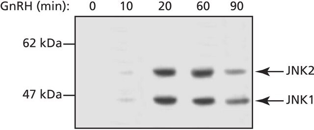
Western Blotting Analysis (WB): A 1:10,000 dilution of this antibody detected EGFR phosphorylated on Tyrosine 1068 in lysate from over-night serum starved A431 cells treated with EGF (50 ng/mL; 10 min.).
Tested Applications
Western Blotting Analysis: A 1:10,000 dilution from a representative lot detected Phospho-EGFR (Tyr1068) in lysate from HeLa cells overnight serum starved and treated with EGF ( (50 ng/mL; 10 min.).
Affinity Binding Assay: A representative lot of this antibody bound Phospho-EGF Receptor-(Tyr1068) peptide with a KD of 1.1 x 10⁻⁶ in an affinity binding assay.
Flow Cytometry Analysis: 1 µg from a representative lot detected Phospho-EGFR (Tyr1068) in one million A431 cells treated with EGF (100 ng/mL; 15 min.).
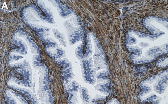
Immunohistochemistry (Paraffin) Analysis: A 1:100 dilution from a representative lot detected Phospho-EGFR (Tyr1068) in human cervical cancer tissue sections.
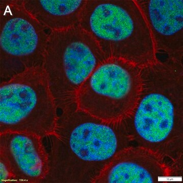
Immunocytochemistry Analysis: A 1:100 dilution from a representative lot detected Phospho-EGFR (Tyr1068) in A431 cells treated with EGF (200 ng/mL; 10 min.).
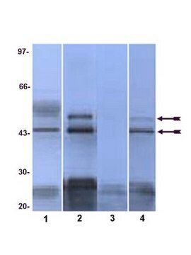
Peptide Inhibition Assay: Target band detection in a lysate from A431 cells treated with EGF (50 ng/mL; 10 min) was prevented by preblocking of a representative lot with the immunogen phosphopeptide, but not the corresponding non-phosphopeptide.
Note: Actual optimal working dilutions must be determined by end user as specimens, and experimental conditions may vary with the end user.
Target description
Physical form
Reconstitution
Storage and Stability
Other Notes
Legal Information
Disclaimer
Not finding the right product?
Try our Product Selector Tool.
Storage Class Code
11 - Combustible Solids
WGK
WGK 1
Flash Point(F)
Not applicable
Flash Point(C)
Not applicable
Choose from one of the most recent versions:
Certificates of Analysis (COA)
Don't see the Right Version?
If you require a particular version, you can look up a specific certificate by the Lot or Batch number.
Already Own This Product?
Find documentation for the products that you have recently purchased in the Document Library.
Our team of scientists has experience in all areas of research including Life Science, Material Science, Chemical Synthesis, Chromatography, Analytical and many others.
Contact Technical Service


