Skip to Content
Merck
All Photos(1)

Documents

04-1624

Sigma-Aldrich

Anti-Centrin Antibody, clone 20H5

clone 20H5, from mouse

Synonym(s):

Caltractin isoform 2, EF-hand protein, calcium binding protein, centrin 1, centrin, EF-hand protein, 1

Sign Into View Organizational & Contract Pricing


About This Item

UNSPSC Code:
12352203
eCl@ss:
32160702
NACRES:
NA.41

biological source

mouse

Quality Level

antibody form

purified immunoglobulin

antibody product type

primary antibodies

clone

20H5, monoclonal

species reactivity

rat, human, mouse

technique(s)

immunocytochemistry: suitable
immunoprecipitation (IP): suitable
western blot: suitable

isotype

IgG2aκ

NCBI accession no.

UniProt accession no.

shipped in

wet ice

target post-translational modification

unmodified

Gene Information

human ... CETN1(1068)

General description

Centrin is a key regulator of centriole duplication. This protein is localized to the centrosome of interphase cells. During mitosis, centrin redistributes to the spindle poles, consistent with dynamic changes of the centrosome during the cell cycle. Current research suggests centrin exerts spatial and temporal control over centriole duplication through its role as a component of centriole precursor structures and through periodic cell-cycle specific changes in its abundance.

Specificity

This antibody recognizes the C-terminus of Centrin.

Immunogen

Epitope: C-terminus
Recombinant protein corresponding to the C-terminus of Chlamydomonas Centrin.

Application

Anti-Centrin Antibody, clone 20H5 is a Mouse Monoclonal Antibody for detection of Centrin also known as Caltractin isoform 2, EF-hand protein & has been validated in WB, IP, ICC.
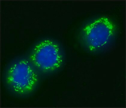
Immunocytochemistry Analysis: Manandhar, G., et al. (1999). Cell Motil Cytoskeleton. 43(2):137-144.
Research Category
Epigenetics & Nuclear Function
Research Sub Category
Cell Cycle, DNA Replication & Repair

Quality

Evaluated by Western Blot in HeLa cell lysate.

Western Blot Analysis: 1 µg/ml of this antibody detected Centrin on 10 µg of HeLa cell lysate.

Target description

~ 20 kDa

Physical form

Format: Purified
Protein G Purified
Purified mouse monoclonal IgG2aκ in buffer containing 0.1 M Tris-Glycine (pH 7.4, 150 mM NaCl) with 0.05% sodium azide.

Storage and Stability

Stable for 1 year at 2-8°C from date of receipt.

Analysis Note

Control
HeLa cell lysate

Other Notes

Concentration: Please refer to the Certificate of Analysis for the lot-specific concentration.

Disclaimer

Unless otherwise stated in our catalog or other company documentation accompanying the product(s), our products are intended for research use only and are not to be used for any other purpose, which includes but is not limited to, unauthorized commercial uses, in vitro diagnostic uses, ex vivo or in vivo therapeutic uses or any type of consumption or application to humans or animals.

Not finding the right product?  

Try our Product Selector Tool.

Storage Class Code

12 - Non Combustible Liquids

WGK

WGK 1

Flash Point(F)

Not applicable

Flash Point(C)

Not applicable


Certificates of Analysis (COA)

Search for Certificates of Analysis (COA) by entering the products Lot/Batch Number. Lot and Batch Numbers can be found on a product’s label following the words ‘Lot’ or ‘Batch’.

Already Own This Product?

Find documentation for the products that you have recently purchased in the Document Library.

Visit the Document Library

Customers Also Viewed

G Manandhar et al.
Cell motility and the cytoskeleton, 43(2), 137-144 (1999-06-24)
Centrosome reduction during mouse spermiogenesis has been studied by immunofluorescent microscopy using anticentrin antibody (20H5) and TEM. Centrin is detected as two spots in round spermatids, corresponding to a pair of centrioles. In elongating spermatids, centrin spots colocalize with the
Rachael A McCloy et al.
Cell cycle (Georgetown, Tex.), 13(9), 1400-1412 (2014-03-15)
Entry and progression through mitosis has traditionally been linked directly to the activity of cyclin-dependent kinase 1 (Cdk1). In this study we utilized low doses of the Cdk1-specific inhibitor, RO3306 from early G 2 phase onwards. Addition of low doses
Stress-induced localization of HSPA6 (HSP70B') and HSPA1A (HSP70-1) proteins to centrioles in human neuronal cells.
Khalouei, S; Chow, AM; Brown, IR
Cell Stress & Chaperones null
Mi Young Seo et al.
PloS one, 10(9), e0138905-e0138905 (2015-09-26)
A procentriole is assembled next to the mother centriole during S phase and remains associated until M phase. After functioning as a spindle pole during mitosis, the mother centriole and procentriole are separated at the end of mitosis. A close
Chia-Yih Wang et al.
Molecular and cellular biology, 33(3), 476-484 (2012-11-21)
Steroidogenic factor 1 (SF-1 or NR5A1) is a nuclear receptor that controls adrenogenital cell growth and differentiation. Adrenogenital primordial cells from SF-1 knockout mice die of apoptosis, but the mechanism by which SF-1 regulates cell survival is not entirely clear.

Our team of scientists has experience in all areas of research including Life Science, Material Science, Chemical Synthesis, Chromatography, Analytical and many others.

Contact Technical Service