Skip to Content
MilliporeSigma
All Photos(3)

Key Documents

T1325

Sigma-Aldrich

Anti-Phosphotyrosine antibody produced in rabbit

0.5 mg/mL, affinity isolated antibody, buffered aqueous solution

Synonym(s):

Anti-p-Tyr

Sign Into View Organizational & Contract Pricing


About This Item

MDL number:
UNSPSC Code:
12352203
NACRES:
NA.44

biological source

rabbit

conjugate

unconjugated

antibody form

affinity isolated antibody

antibody product type

primary antibodies

clone

polyclonal

form

buffered aqueous solution

concentration

0.5 mg/mL

technique(s)

indirect immunofluorescence: 3-6 μg/mL using A431 stimulated by human EGF.
western blot: 0.3-0.6 μg/mL using total cell extract of A431 stimulated by human EGF

shipped in

dry ice

storage temp.

−20°C

target post-translational modification

phosphorylation (pTyr)

Specificity

Anti-Phosphotyrosine recognizes tyrosine phosphorylated proteins.

Immunogen

phospho-L-tyrosine conjugated to KLH

Application

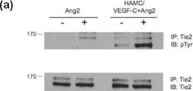
Anti-Phosphotyrosine antibody is suitable for use in indirect immunofluorescence (3-6 μg/mL using A431 stiμLated by human EGF) and immunoblot (using U2OS Dr1 cells ). The antibody may also be used for immunoprecipitation (1:40 using rat lumbar and thoracic lysates) and immunocytochemistry.

Biochem/physiol Actions

Protein phosphorylation is the most abundant among post-translational modifications of cellular proteins, regulating intracellular signal transduction pathways in every living cell. Tyrosine, serine and threonine are the major amino acids that are phosphorylated in proteins. Antibodies specific for phosphotyrosine are essential tools for the characterization of tyrosine phosphorylation in many signal transduction pathways.

Physical form

Solution in 0.01 M phosphate buffered saline, pH 7.4, and 1% BSA, containing 15 mM sodium azide.

Storage and Stability

For extended storage, freeze in working aliquots. Repeated freezing and thawing is not recommended. Storage in "frost-free" freezers is not recommended. If slight turbidity occurs upon prolonged storage, clarify the solution by centrifugation before use. Working dilution samples should be discarded if not used within 12 hours.

Disclaimer

Unless otherwise stated in our catalog or other company documentation accompanying the product(s), our products are intended for research use only and are not to be used for any other purpose, which includes but is not limited to, unauthorized commercial uses, in vitro diagnostic uses, ex vivo or in vivo therapeutic uses or any type of consumption or application to humans or animals.

Not finding the right product?  

Try our Product Selector Tool.

Storage Class Code

12 - Non Combustible Liquids

WGK

nwg

Flash Point(F)

Not applicable

Flash Point(C)

Not applicable


Certificates of Analysis (COA)

Search for Certificates of Analysis (COA) by entering the products Lot/Batch Number. Lot and Batch Numbers can be found on a product’s label following the words ‘Lot’ or ‘Batch’.

Already Own This Product?

Find documentation for the products that you have recently purchased in the Document Library.

Visit the Document Library

A Wiedłocha et al.
Molecular and cellular biology, 16(1), 270-280 (1996-01-01)
U2OS Dr1 cells, originating from a human osteosarcoma, are resistant to the intracellular action of diphtheria toxin but contain toxin receptors on their surfaces. These cells do not have detectable amounts of fibroblast growth factor receptors. When these cells were
Ying Xu et al.
Carcinogenesis, 37(2), 215-222 (2016-01-01)
Dysregulated expression of epidermal growth factor receptor (EGFR) has been implicated in many cancer events, while peroxisome proliferator-activated receptor γ (PPARγ) negatively regulates cancer progression. The molecular mechanism of EGFR interaction with PPARγ is still unclear. Here, we found that
Rafael S Demarco et al.
Methods (San Diego, Calif.), 68(1), 218-227 (2014-05-07)
The process of spermatogenesis in Drosophila melanogaster provides a powerful model system to probe a variety of developmental and cell biological questions, such as the characterization of mechanisms that regulate stem cell behavior, cytokinesis, meiosis, and mitochondrial dynamics. Classical genetic
Shuangsong Hong et al.
The Journal of biological chemistry, 279(28), 29341-29350 (2004-05-05)
Diabetic neuropathy is a common form of peripheral neuropathy, yet the mechanisms responsible for pain in this disease are poorly understood. Alterations in the expression and function of voltage-gated tetrodotoxin-resistant (TTX-R) sodium channels have been implicated in animal models of
Michael Bachmann et al.
Journal of cell science, 135(7) (2022-03-29)
Integrin-mediated adhesions are convergence points for multiple signaling pathways. Their inner structure and diverse functions can be studied with super-resolution microscopy. Here, we examined the spatial organization within focal adhesions by analyzing several adhesion proteins with structured illumination microscopy (SIM).

Our team of scientists has experience in all areas of research including Life Science, Material Science, Chemical Synthesis, Chromatography, Analytical and many others.

Contact Technical Service