SCR077
Fluorescent Mouse ES/iPS Cell Characterization Kit
This Fluorescent Mouse ES/iPS Cell Characterization Kit contains a range of sensitive tools for the phenotypic assessment of the pluripotent status of mouse Embryonic stem & induced pluripotent Stem cells.
Sign Into View Organizational & Contract Pricing
All Photos(5)
About This Item
UNSPSC Code:
12161503
eCl@ss:
32161000
NACRES:
NA.84
Recommended Products
General description
Mouse embryonic stem (mES) cells are pluripotent cells derived from the inner cell mass of pre-implantation blastocysts. Mouse induced pluripotent (miPS) cells are pluripotent cells generated by reprogramming mouse somatic cells using four transcription factors, Oct-4, Klf-4, Sox-2, and c-Myc, or their variants. Both mESC and miPSC can self-renew and have the ability to generate all three germ layers: ectoderm, mesoderm, and endoderm. In vitro, mESC/iPSC are normally maintained and propagated on mouse fibroblast feeders or in feeder-free conditions in media containing LIF/ESGRO. However, spontaneous differentiation may occur in subpopulations of cells. Several pluripotent markers are commonly used to distinguish pluripotent mESC/iPSC from differentiated cells.
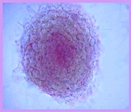
EMD Millipore’s Fluorescent Mouse ES/iPS Cell Characterization Kit contains a range of sensitive tools for the phenotypic assessment of the pluripotent status of mouse ES/iPS cells. Included in the kit is an enzymatic assay to measure alkaline phosphatase activity in the cells along with validated directly conjugated antibodies to pluripotent transcription factors, Oct-4, Sox-2 and DPPA-2 and cell surface epitope SSEA-1 to enable rapid immunocytochemical marker analysis. The Dapi nuclear dye is conveniently included to aid in cell quantification. While the expression levels of pluripotent markers are expected to be diminished upon differentiation, each possess specific expression kinetics.
EMD Millipore’s Fluorescent Mouse ES/iPS Cell Characterization Kit contains a range of sensitive tools for the phenotypic assessment of the pluripotent status of mouse ES/iPS cells. Included in the kit is an enzymatic assay to measure alkaline phosphatase activity in the cells along with validated directly conjugated antibodies to pluripotent transcription factors, Oct-4, Sox-2 and DPPA-2 and cell surface epitope SSEA-1 to enable rapid immunocytochemical marker analysis. The Dapi nuclear dye is conveniently included to aid in cell quantification. While the expression levels of pluripotent markers are expected to be diminished upon differentiation, each possess specific expression kinetics.
Specificity
Cross Reactivty
None
None
Application
Research Category
Stem Cell Research
Stem Cell Research
Research Sub Category
Pluripotent & Early Differentiation
Pluripotent & Early Differentiation
This Fluorescent Mouse ES/iPS Cell Characterization Kit contains a range of sensitive tools for the phenotypic assessment of the pluripotent status of mouse Embryonic stem & induced pluripotent Stem cells.
Components
1. Fast Red Violet solution (Part No. 90239): One 15 mL bottle.
2. Napthol AS-BI phosphate solution (Part No. 90234). One 15 mL bottle.
3. Mouse anti-Oct-4 (POU5f1), clone 7F9.2, Alexa Fluor™ 488 conjugate (Part No. MAB4419A4-50UL). One vial containing 50 µL of 0.5 mg/mL conjugated monoclonal antibody.
4. Mouse anti-Sox-2, clone 10H9.1, Cy3 conjugate (Part No. MAB4423C3-50UL). One vial containing 50 µL of 0.5 mg/mL conjugated monoclonal antibody.
5. Mouse anti-DPPA-2, clone 6C1.2, Alexa Fluor 488 conjugate (Part No. MAB4356A4-50UL). One vial containing 50 µL of 0.5 mg/mL conjugated monoclonal antibody.
5. Mouse anti-SSEA-1, clone MC-480, Cy3 conjugate (Part No. MAB4301C3-50UL). One vial containing 50 µL of 0.5 mg/mL conjugated monoclonal antibody.
8. DAPI, 100 µL (Part No. 90229). One vial containing 100 µL volume.
Quality
Conjugated antibodies and alkaline phosphatase reagents have been validated on mouse ES and STEMCCA derived mouse ips cells.
Physical form
Purified conjugated mouse monoclonal antibodies are stored in PBS with 0.1% sodium azide and 15mg/ml BSA.
Storage and Stability
Store at 2-8°C until expiration date of kit. Do not freeze or expose to elevated temperatures. Discard any remaining reagents after the expiration date.
Legal Information
ALEXA FLUOR is a trademark of Life Technologies
Signal Word
Warning
Hazard Statements
Precautionary Statements
Hazard Classifications
Carc. 2 - Eye Irrit. 2 - Skin Irrit. 2
Storage Class Code
10 - Combustible liquids
Certificates of Analysis (COA)
Search for Certificates of Analysis (COA) by entering the products Lot/Batch Number. Lot and Batch Numbers can be found on a product’s label following the words ‘Lot’ or ‘Batch’.
Already Own This Product?
Find documentation for the products that you have recently purchased in the Document Library.
Yanbo Zhu et al.
International journal of biological sciences, 16(11), 1861-1875 (2020-05-14)
Induced pluripotent stem cells (iPSCs), derived from reprogramming of somatic cells by a cocktail of transcription factors, have the capacity for unlimited self-renewal and the ability to differentiate into all of cell types present in the body. iPSCs may have
Cong Wang et al.
Theranostics, 10(1), 353-370 (2020-01-07)
Background: Long non-coding RNAs (lncRNAs) constitute an important component of the regulatory apparatus that controls stem cell pluripotency. However, the specific mechanisms utilized by these lncRNAs in the control of pluripotency are not fully characterized. Methods: We utilized a RNA
Zhonghua Du et al.
Genome biology, 22(1), 233-233 (2021-08-21)
A specific 3-dimensional intrachromosomal architecture of core stem cell factor genes is required to reprogram a somatic cell into pluripotency. As little is known about the epigenetic readers that orchestrate this architectural remodeling, we used a novel chromatin RNA in
Zhonghua Du et al.
Scientific data, 5, 180255-180255 (2018-11-21)
Pluripotent stem cells hold great investigative potential for developmental biology and regenerative medicine. Recent studies suggest that long noncoding RNAs (lncRNAs) may function as key regulators of the maintenance and the lineage differentiation of stem cells. However, the underlying mechanisms
Lin Jia et al.
Nucleic acids research, 48(7), 3935-3948 (2020-02-15)
Formation of a pluripotency-specific chromatin network is a critical event in reprogramming somatic cells into pluripotent status. To characterize the regulatory components in this process, we used 'chromatin RNA in situ reverse transcription sequencing' (CRIST-seq) to profile RNA components that
Our team of scientists has experience in all areas of research including Life Science, Material Science, Chemical Synthesis, Chromatography, Analytical and many others.
Contact Technical Service


