MABF959
Anti-CD47 Antibody, clone C5/D5
clone C5/D5, from mouse
Synonym(s):
Leukocyte surface antigen CD47, Antigenic surface determinant protein OA3, CD47, IAP, Integrin-associated protein, Protein MER6
About This Item
Recommended Products
biological source
mouse
Quality Level
antibody form
purified immunoglobulin
antibody product type
primary antibodies
clone
C5/D5, monoclonal
species reactivity
human
technique(s)
flow cytometry: suitable
isotype
IgG1κ
NCBI accession no.
UniProt accession no.
shipped in
ambient
target post-translational modification
unmodified
Gene Information
human ... CD47(961)
Related Categories
General description
Specificity
Immunogen
Application
Flow Cytometry Analysis: A representative lot detected the expression of exogenously transfected human CD47 on the surface of CaCO2 cells (Liu, Y., et al. (2001). J. Biol. Chem. 276(43):40156-40166).
Flow Cytometry Analysis: Representative lots detected human polymorphonuclear (PMN) neutrophils surface CD47 immunoreactivity, which was upregulated after fMLP stimulation (Liu, Y., et al. (2001). J. Biol. Chem. 276(43):40156-40166; Parkos, C.A., et al. (1996). J. Cell Biol. 132(3):437-450).
Immunoaffinity Purification: A representative lot was immobilized on resins and employed for affinity purification of CD47 from HT29 (subclone Cl 19.A) lysate (Parkos, C.A., et al. (1996). J. Cell Biol. 132(3):437-450).
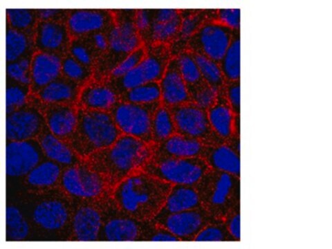
Immunocytochemistry Analysis: A representative lot immunostained the basolateral surfaces of methanol-fixed CaCO2 monolayers expressing exogenously transfected human CD47 by fluorescent immunocytochemistry (Liu, Y., et al. (2001). J. Biol. Chem. 276(43):40156-40166).
Immunocytochemistry Analysis: A representative lot immunostained the basolateral membrane of paraformaldehyde-fixed T84 human intestinal epithelial cell monolayer by fluorescent immunocytochemistry (Parkos, C.A., et al. (1996). J. Cell Biol. 132(3):437-450).
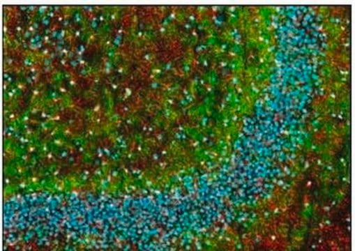
Immunofluorescence Analysis: A representative lot immunostained the colonic crypts and lamina propria leukocytes at the basal and lateral, but not apical, aspects of the epithelium in paraformaldehyde-fixed frozen human colon tissue sections by fluorescent immunohistochemistry (Parkos, C.A., et al. (1996). J. Cell Biol. 132(3):437-450).
Immunoprecipitation Analysis: A representative lot immunoprecipitated ~60-65 kDa glycosylated CD47 from T84 human intestinal epithelial cell lysate. Target band size reduced to ~35 kDa following deglycosylation by N glycosidase F treatment (Parkos, C.A., et al. (1996). J. Cell Biol. 132(3):437-450).
Neutralizing Analysis: Representative lots caused a delay of human polymorphonuclear (PMN) neutrophils migration across polarized monolayers of T84 human intestinal epithelial cells in a bidirectional fashion without affecting PMN neutrophils or T84 adhesion. Tyrosine phosphorylation inhibitor genistein prevented delay by clone C5/D5 (Liu, Y., et al. (2001). J. Biol. Chem. 276(43):40156-40166; Parkos, C.A., et al. (1996). J. Cell Biol. 132(3):437-450).
Inflammation & Immunology
Quality
Flow Cytometry Analysis: 0.4 µL of this antibody detected CD47 surface expression on the gated lymphocytes population among one million human PBMCs.
Target description
Physical form
Storage and Stability
Handling Recommendations: Upon receipt and prior to removing the cap, centrifuge the vial and gently mix the solution. Aliquot into microcentrifuge tubes and store at -20°C. Avoid repeated freeze/thaw cycles, which may damage IgG and affect product performance.
Other Notes
Disclaimer
Not finding the right product?
Try our Product Selector Tool.
Storage Class Code
12 - Non Combustible Liquids
WGK
WGK 2
Flash Point(F)
Not applicable
Flash Point(C)
Not applicable
Certificates of Analysis (COA)
Search for Certificates of Analysis (COA) by entering the products Lot/Batch Number. Lot and Batch Numbers can be found on a product’s label following the words ‘Lot’ or ‘Batch’.
Already Own This Product?
Find documentation for the products that you have recently purchased in the Document Library.
Our team of scientists has experience in all areas of research including Life Science, Material Science, Chemical Synthesis, Chromatography, Analytical and many others.
Contact Technical Service


