Skip to Content
MilliporeSigma
All Photos(1)

Key Documents

MABC943

Sigma-Aldrich

Anti-OAS1 Antibody, clone 1D11.1

clone 1D11.1, from mouse

Synonym(s):

2′-5′-oligoadenylate synthase 1, (2-5′)oligo(A) synthase 1, 2-5A synthase 1, E18/E16, p46/p42 OAS

Sign Into View Organizational & Contract Pricing


About This Item

UNSPSC Code:
12352203
eCl@ss:
32160702
NACRES:
NA.43

biological source

mouse

Quality Level

antibody form

purified antibody

antibody product type

primary antibodies

clone

1D11.1, monoclonal

species reactivity

human

technique(s)

western blot: suitable

isotype

IgG2aκ

NCBI accession no.

UniProt accession no.

target post-translational modification

unmodified

Gene Information

human ... OAS1(4938)

General description

2′-5′-oligoadenylate synthase 1 (EC 2.7.7.84; UniProt P00973; also known as (2-5′)oligo(A) synthase 1, 2-5A synthase 1, E18/E16, p46/p42 OAS) is encoded by the OAS1 (also known as OIAS, OIASI) gene (Gene ID 4938) in human. Pattern recognition receptors (PRRs) recognize pathogen-associated molecular patterns (PAMPs), including pathogens-derived double-stranded RNA and DNA (dsRNA and dsDNA). The 2′-5′ oligoadenylate synthetases (OAS) are interferon-induced cytoplasmic PRRs that recognize virally produced dsRNA and initiate RNA destabilization through activation of RNase L within infected cells. Upon dsRNA binding, OAS enzymes generate the second messenger 2’-5’ oligoadenylate (2-5An; n = 2 to 19), resulting in the dimerization and activation of the latent ribonuclease (RNase L) that degrades host and viral mRNAs. In primates, the OAS family consists of OAS1, OAS2, OAS3, and the catalytically inactive OASL, while 12 rodent Oas genes have been identified, eight of which are OAS1 paralogs.

Specificity

Clone 1D11.1 was raised against a recombinant fragment derived from the spliced isoform p46 (E18; Uniprot P00973-1), cross-reactivity toward the p41 (E16; 3-9), p48 (9-2), and p44 spliced isoforms is possible, but has not been determined.

Immunogen

Epitope: Internal (C-terminal half).
GST-tagged recombinant internal fragment from the C-terminal half of human OAS1.

Application

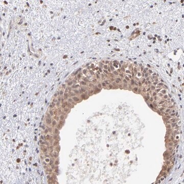
Anti-OAS1 Antibody, clone 1D11.1 is an antibody against OAS1 for use in Western Blotting.
Research Category
Apoptosis & Cancer
Research Sub Category
RNA Metabolism & Binding Proteins

Quality

Evaluated by Western Blotting in lysate of IFN gamma-treated HeLa cells.

Western Blotting Analysis: A 1:500 dilution of this antibody detected OAS1 in 10 µg of lysate from IFN gamma-treated HeLa cells.

Target description

~50 kDa observed. 46.03 kDa (isoform p46; E18), 41.74 kDa (isoform p41; E16; 3-9), 47.41 kDa (isoform p48; 9-2), and 43.94 kDa (isoform p44) calculated.

Physical form

Format: Purified
Protein G Purified
Purified mouse monoclonal IgG2aκ antibody in buffer containing 0.1 M Tris-Glycine (pH 7.4), 150 mM NaCl with 0.05% sodium azide.

Storage and Stability

Stable for 1 year at 2-8°C from date of receipt.

Other Notes

Concentration: Please refer to lot specific datasheet.

Disclaimer

Unless otherwise stated in our catalog or other company documentation accompanying the product(s), our products are intended for research use only and are not to be used for any other purpose, which includes but is not limited to, unauthorized commercial uses, in vitro diagnostic uses, ex vivo or in vivo therapeutic uses or any type of consumption or application to humans or animals.

Not finding the right product?  

Try our Product Selector Tool.

Storage Class Code

12 - Non Combustible Liquids

WGK

WGK 1

Flash Point(F)

Not applicable

Flash Point(C)

Not applicable


Certificates of Analysis (COA)

Search for Certificates of Analysis (COA) by entering the products Lot/Batch Number. Lot and Batch Numbers can be found on a product’s label following the words ‘Lot’ or ‘Batch’.

Already Own This Product?

Find documentation for the products that you have recently purchased in the Document Library.

Visit the Document Library

Runhong Zhou et al.
Journal of virology, 92(6) (2017-12-22)
Although it has been shown that some mannose-binding lectins (MBLs) exhibit significant activity against HIV infection, little is known about whether N-acetylgalactosamine (GalNAc)-binding lectins have the ability to inhibit HIV infection. Here, we demonstrate that a soybean-derived lectin (SBL) with

Our team of scientists has experience in all areas of research including Life Science, Material Science, Chemical Synthesis, Chromatography, Analytical and many others.

Contact Technical Service