Skip to Content
MilliporeSigma
All Photos(2)

Key Documents

ABT332

Sigma-Aldrich

Anti-ZEB2 Antibody

from rabbit, purified by affinity chromatography

Synonym(s):

Zinc finger E-box binding homeobox 2, Smad interacting-protein 1

Sign Into View Organizational & Contract Pricing


About This Item

UNSPSC Code:
12352203
eCl@ss:
32160702
NACRES:
NA.41

biological source

rabbit

Quality Level

antibody form

affinity isolated antibody

antibody product type

primary antibodies

clone

polyclonal

purified by

affinity chromatography

species reactivity

human

technique(s)

immunocytochemistry: suitable
immunofluorescence: suitable
western blot: suitable

UniProt accession no.

shipped in

wet ice

target post-translational modification

unmodified

Gene Information

human ... ZEB2(9839)

General description

Zinc finger E-box-binding homeobox 2 (ZEB2), also known as Smad-interacting protein 1 (SIP1, SMADIP1) and Zinc finger homeobox protein 1b (ZFHX1B, ZFX1B), is a transcriptional inhibitor that binds to DNA sequence 5′-CACCT-3′ in different promoters and interacts with activated SMADs. ZEB2 has been shown to repress the transcription of the E-cadherin gene and induce epithelial-mesenchymal transition, a genetic program controlling cell migration during embryonic development and wound healing in vitro. It is suggested that ZEB2 contributes to tumor progression since it’s been demonstrated to protect cells from DNA damage-induced apoptosis. Recent studies have shown that ZEB2 is often observed in the cytoplasm in numerous cancer tissues, implying that its localization may be regulated in normal and tumor tissues. Mutations in this gene are also associated with Mowat-Wilson syndrome (MWIS), also known as Hirschsprung disease-mental syndrome.

Immunogen

KLH-conjugated linear peptide corresponding to human ZEB2.

Application

Detect ZEB2 using this rabbit polyclonal antibody, Anti-ZEB2 Antibody validated for use in western blotting, ICC & Immunofluorescence.
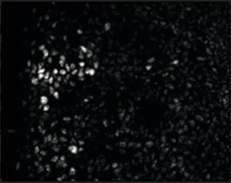
Immunocytochemistry Analysis: 20 µg/mL from a representative lot detected ZEB2 in Jurkat cells.

Immunofluorescence Analysis: 20 μg/mL from a representative lot detected ZEB2 in Jurkat cells.
Research Category
Cell Structure
Research Sub Category
Adhesion (CAMs)

Quality

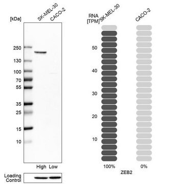
Evaluated by Western Blotting in Jurkat cell lysate.

Western Blotting Analysis: 2 µg/mL of this antibody detected ZEB2 in 15 µg of Jurkat cell lysate.

Target description

~200 kDa observed. Uncharacterized bands may appear in some lysate(s). Uniprot describes 2 isoforms at ~134 kDa and ~136 kDa The calculated molecular weight of this protein is 136 kDa, but can be observed at ~200 kDa due to post-translational modification.

Physical form

Antigen Affinity Purified
Purified rabbit polyclonal in buffer containing PBS with up to 0.1% sodium azide.

Storage and Stability

Stable for 1 year at 2-8°C from date of receipt.

Analysis Note

Control
Jurkat cell lysate

Other Notes

Concentration: Please refer to the Certificate of Analysis for the lot-specific concentration.

Disclaimer

Unless otherwise stated in our catalog or other company documentation accompanying the product(s), our products are intended for research use only and are not to be used for any other purpose, which includes but is not limited to, unauthorized commercial uses, in vitro diagnostic uses, ex vivo or in vivo therapeutic uses or any type of consumption or application to humans or animals.

Not finding the right product?  

Try our Product Selector Tool.

Storage Class Code

10 - Combustible liquids

WGK

WGK 2

Flash Point(F)

Not applicable

Flash Point(C)

Not applicable


Certificates of Analysis (COA)

Search for Certificates of Analysis (COA) by entering the products Lot/Batch Number. Lot and Batch Numbers can be found on a product’s label following the words ‘Lot’ or ‘Batch’.

Already Own This Product?

Find documentation for the products that you have recently purchased in the Document Library.

Visit the Document Library

Olga Ilina et al.
Nature cell biology, 22(9), 1103-1115 (2020-08-26)
Plasticity of cancer invasion and metastasis depends on the ability of cancer cells to switch between collective and single-cell dissemination, controlled by cadherin-mediated cell-cell junctions. In clinical samples, E-cadherin-expressing and -deficient tumours both invade collectively and metastasize equally, implicating additional
Jonathan M Goodwin et al.
Molecular cell, 55(3), 436-450 (2014-07-22)
The serine/threonine kinase LKB1 is a tumor suppressor whose loss is associated with increased metastatic potential. In an effort to define biochemical signatures of metastasis associated with LKB1 loss, we discovered that the epithelial-to-mesenchymal transition transcription factor Snail1 was uniquely

Our team of scientists has experience in all areas of research including Life Science, Material Science, Chemical Synthesis, Chromatography, Analytical and many others.

Contact Technical Service