Skip to Content
MilliporeSigma
All Photos(1)

Key Documents

05-598

Sigma-Aldrich

Anti-phospho-Histone H3 (Ser10) Antibody, clone RR002

clone RR002, Upstate®, from mouse

Synonym(s):

H3 histone, family 3A, H3S10P, Histone H3 (phospho S10), H3 histone, family 3A, H3 histone, family 3B, H3 histone, family 3B (H3.3B)

Sign Into View Organizational & Contract Pricing


About This Item

UNSPSC Code:
12352203
eCl@ss:
32160702
NACRES:
NA.41

biological source

mouse

Quality Level

antibody form

purified immunoglobulin

antibody product type

primary antibodies

clone

RR002, monoclonal

species reactivity

vertebrates, nematode, Caenorhabditis elegans, human

manufacturer/tradename

Upstate®

technique(s)

immunocytochemistry: suitable
immunohistochemistry: suitable
western blot: suitable

isotype

IgG

suitability

not suitable for ELISA applications

NCBI accession no.

UniProt accession no.

shipped in

wet ice

target post-translational modification

phosphorylation (pSer10)

Gene Information

human ... HIST1H3F(8968)

General description

Histone H3 is one of the five main histone proteins involved in the structure of chromatin in eukaryotic cells. Featuring a main globular domain and a long N-terminal tail, H3 is involved with the structure of the nucleosomes of the ′beads on a string′ structure. The N-terminal tail of histone H3 protrudes from the globular nucleosome core and can undergo several different types of epigenetic modifications that influence cellular processes. These modifications include the covalent attachment of methyl or acetyl groups to lysine and arginine amino acids and the phosphorylation of serine or threonine.

Specificity

Broad species cross-reactivity expected due to sequence conservation.
This antibody recognizes Histone H3 phosphorylated at Serine 10.

Immunogen

Peptide (ARK[pS]TGGKAPRKQL-C) corresponding to amino acids 7-20 of human histone H3 with a c-terminal cyteine added for conjugation purposes.

Application

Anti-phospho-Histone H3 (Ser10) Antibody, clone RR002 is a Mouse Monoclonal Antibody for detection of Histone H3 phosphorylated at serine 10. This purified mAb, also known as Anti-H3S10p, is published in peer reviewed journals and is validated in ICC, IHC, WB.
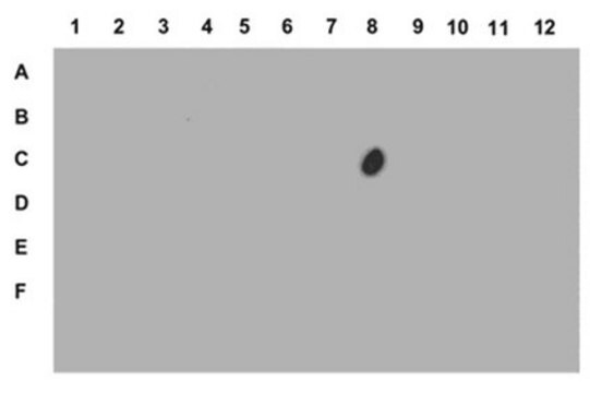
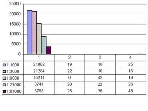
ELISA Analysis:
This antibody showed no immuno-reactivity with an unphosphorylated Histone H3 peptide (residues 7-20), nor with other histone phosphopeptides.

Immunohistochemistry:
A 1:1000 dilution of this antibody detected mitotic cells in C. elegans embryo sections as shown by an independent laboratory.

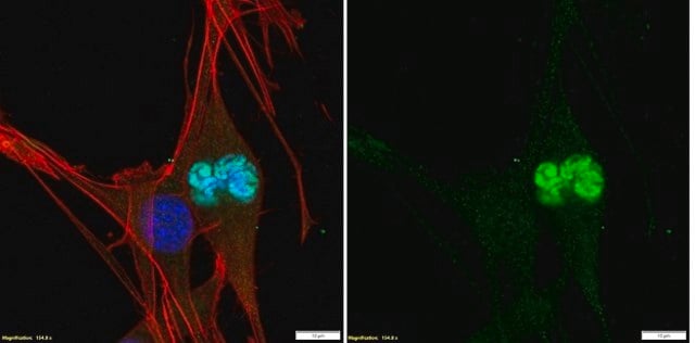
Immunocytochemistry:
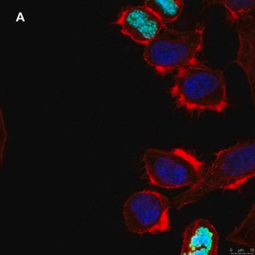
This lot showed positive staining of mitotic cells in cultures of HeLa cells undergoing exponential growth

Quality

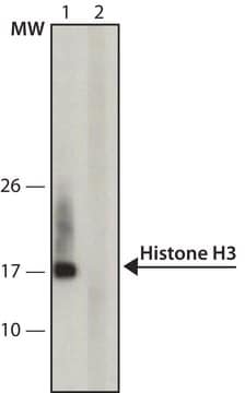
Routinely evaluated by western blot on acid extracted proteins from colcemid-arrested HeLa cells.

Western Blot Analysis:
0.005-0.025 μg/mL of this lot detected phosphorylated Histone H3 in acid extracted proteins from colcemid-arrested HeLa cells.

Target description

17 kDa

Physical form

Format: Purified
Purified mouse IgG in buffer containing 0.1M Tris Glycine, 0.15M NaCl, 0.05% Sodium Azide, pH 7.4.

Analysis Note

Control
UV-treated 293 cell extracts, UV-treated HeLa cell extracts, breast cancer tissue, HEPG2 cell extrats.

Other Notes

Concentration: Please refer to the Certificate of Analysis for the lot-specific concentration.

Legal Information

UPSTATE is a registered trademark of Merck KGaA, Darmstadt, Germany

Not finding the right product?  

Try our Product Selector Tool.

Storage Class Code

10 - Combustible liquids

WGK

WGK 1


Certificates of Analysis (COA)

Search for Certificates of Analysis (COA) by entering the products Lot/Batch Number. Lot and Batch Numbers can be found on a product’s label following the words ‘Lot’ or ‘Batch’.

Already Own This Product?

Find documentation for the products that you have recently purchased in the Document Library.

Visit the Document Library

Protein phosphatase 2A activity affects histone H3 phosphorylation and transcription in Drosophila melanogaster
Nowak, S. J., et al
Molecular and cellular biology, 23, 6129-6138 (2003)
Joao P Lopes et al.
Aging cell, 9(1), 64-77 (2009-11-10)
Cdk5 dysregulation is a major event in the neurodegenerative process of Alzheimer's disease (AD). In vitro studies using differentiated neurons exposed to Abeta exhibit Cdk5-mediated tau hyperphosphorylation, cell cycle re-entry and neuronal loss. In this study we aimed to determine
Functional consequences of cyclin D1/BRCA1 interaction in breast cancer cells.
Kehn, K; Berro, R; Alhaj, A; Bottazzi, ME; Yeh, WI; Klase, Z; Van Duyne, R; Fu, S; Kashanchi, F
Oncogene null
p38-Dependent marking of inflammatory genes for increased NF-kappa B recruitment
Saccani, S., et al
Nature Immunology, 3, 69-75 (2002)
Transmembrane potential of GlyCl-expressing instructor cells induces a neoplastic-like conversion of melanocytes via a serotonergic pathway.
Blackiston, D; Adams, DS; Lemire, JM; Lobikin, M; Levin, M
Disease models & mechanisms null

Our team of scientists has experience in all areas of research including Life Science, Material Science, Chemical Synthesis, Chromatography, Analytical and many others.

Contact Technical Service