Skip to Content
MilliporeSigma
All Photos(1)

Key Documents

05-1324

Sigma-Aldrich

Anti-phospho-Histone H1 Antibody, clone 12D11

culture supernatant, clone 12D11, from mouse

Synonym(s):

H1 histone family, member 5, Histone H1a, histone 1, H1b, histone cluster 1, H1b

Sign Into View Organizational & Contract Pricing


About This Item

UNSPSC Code:
12352203
eCl@ss:
32160702
NACRES:
NA.41

biological source

mouse

Quality Level

antibody form

culture supernatant

antibody product type

primary antibodies

clone

12D11, monoclonal

species reactivity

human, mouse, bovine, vertebrates

technique(s)

immunocytochemistry: suitable
immunohistochemistry: suitable
immunoprecipitation (IP): suitable
western blot: suitable

isotype

IgMκ

NCBI accession no.

UniProt accession no.

shipped in

wet ice

target post-translational modification

phosphorylation (not specified)

Gene Information

bovine ... H1-5(527304)
human ... H1-5(3009)

General description

Histones are highly conserved proteins that serve as the structural scaffold for the organization of nuclear DNA into chromatin. The four core histones, H2A, H2B, H3, and H4, assemble into an octamer (2 molecules of each). Subsequently, 146 base pairs of DNA are wrapped around the octamer, forming a nucleosome. The linker histone, H1, interacts with linker DNA between nucleosomes and functions in the compaction of chromatin into 30nm chromatin fibers and higher order structures.

Specificity

Broad species cross-reactivity is expected, based on sequence homology.
Recognizes phosphorylated Histone H1, Mr 32 kDa.

Immunogen

The immunogen was KLH-conjugated bovine nuclear extract.

Application

Immunohistochemistry: This antibody has been reported by an independent laboratory to detect CDK (cyclin dependent kinase) phosphorylated Histone H1 in benign tissue sections.1

Immunocytochemistry:
This antibody has been demonstrated by an outside laboratory to show positive immunostaining for phosphorylated histone H1 in HUSK (human fetal primary myoblasts) cells fixed with 3.7% paraformaldehyde followed by permeabilization with 0.2% Triton X-100.2

Immunoprecipitation:
This antibody has been demonstrated by an outside laboratory to immunoprecipitate phosphorylated histone H1 using solid phase beads bound to anti-mouse IgM.
Research Category
Epigenetics & Nuclear Function
Research Sub Category
Histones
Use Anti-phospho-Histone H1 Antibody, clone 12D11 (Mouse Monoclonal Antibody) validated in IHC, ICC, IP, WB to detect phospho-Histone H1 also known as Histone H1a, histone 1 H1b.

Quality

Western Blot Analysis:
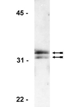
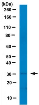
1:1000 dilution of this antibody detected Histone H1 in colcemid-treated HeLa lysate.

Target description

~32 kDa

Physical form

Supplied as 200 μL of culture supernatant with 0.05% sodium azide.

Storage and Stability

Stable for 1 year at -20°C from date of receipt. Upon first thaw, and prior to removing the cap, centrifuge the vial and gently mix the solution. Aliquot into microcentrifuge tubes and store at -20°C. Avoid repeated freeze/thaw cycles, which may damage IgG and affect product performance.

Analysis Note

Control
HeLa cell lysate, treated with colcemid.

Disclaimer

Unless otherwise stated in our catalog or other company documentation accompanying the product(s), our products are intended for research use only and are not to be used for any other purpose, which includes but is not limited to, unauthorized commercial uses, in vitro diagnostic uses, ex vivo or in vivo therapeutic uses or any type of consumption or application to humans or animals.

Not finding the right product?  

Try our Product Selector Tool.

Storage Class Code

12 - Non Combustible Liquids

WGK

WGK 1

Flash Point(F)

Not applicable

Flash Point(C)

Not applicable


Certificates of Analysis (COA)

Search for Certificates of Analysis (COA) by entering the products Lot/Batch Number. Lot and Batch Numbers can be found on a product’s label following the words ‘Lot’ or ‘Batch’.

Already Own This Product?

Find documentation for the products that you have recently purchased in the Document Library.

Visit the Document Library

Growth factors that repress myoblast differentiation sustain phosphorylation of a specific site on histone H1.
Cole, F, et al.
The Journal of Biological Chemistry, 268, 1580-1585 (1993)
Lisa L Hall et al.
The Journal of cell biology, 186(4), 491-507 (2009-08-26)
How XIST RNA strictly localizes across the inactive X chromosome is unknown; however, prophase release of human XIST RNA provides a clue. Tests of inhibitors that mimic mitotic chromatin modifications implicated an indirect role of PP1 (protein phosphatase 1), potentially
Wei-Chung Lai et al.
Scientific reports, 6, 33716-33716 (2016-09-21)
Candida albicans is an opportunistic human fungal pathogen. The ability to switch among multiple cellular forms is key to its pathogenesis. The Dbf4-dependent protein kinase gene CDC7 is conserved due to its role in initiating DNA replication. Because a C.
Nanfei Yang et al.
Cell death and differentiation (2022-11-05)
Beige adipocytes in mammalian white adipose tissue (WAT) can reinforce fat catabolism and energy expenditure. Promoting beige adipocyte biogenesis is a tantalizing tactic for combating obesity and its associated metabolic disorders. Here, we report that a previously unidentified phosphorylation pattern
Yuxiao Chen et al.
Journal of integrative plant biology, 62(11), 1674-1687 (2020-05-30)
Cell division is precisely regulated and highly tissue-specific; studies have suggested that diverse signals in the epidermis, especially the epidermal brassinosteroids (BRs), can regulate root growth. However, the underlying molecular mechanisms that integrate hormonal cues such as BR signaling with

Our team of scientists has experience in all areas of research including Life Science, Material Science, Chemical Synthesis, Chromatography, Analytical and many others.

Contact Technical Service