Skip to Content
Merck
All Photos(1)

Key Documents

223A-1

Sigma-Aldrich

A-1-Antitrypsin Rabbit Polyclonal Antibody

Sign Into View Organizational & Contract Pricing


About This Item

UNSPSC Code:
12352203
NACRES:
NA.41

biological source

rabbit

Quality Level

100
500

conjugate

unconjugated

antibody form

Ig fraction of antiserum

antibody product type

primary antibodies

clone

polyclonal

description

For In Vitro Diagnostic Use in Select Regions (See Chart)

form

buffered aqueous solution

species reactivity

human

packaging

vial of 0.1 mL concentrate (223A-14)
vial of 0.5 mL concentrate (223A-15)
bottle of 1.0 mL predilute (223A-17)
vial of 1.0 mL concentrate (223A-16)
bottle of 7.0 mL predilute (223A-18)

manufacturer/tradename

Cell Marque

technique(s)

immunohistochemistry (formalin-fixed, paraffin-embedded sections): 1:500-1:2000

control

tonsil

shipped in

wet ice

storage temp.

2-8°C

visualization

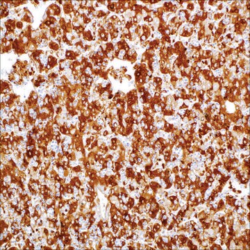
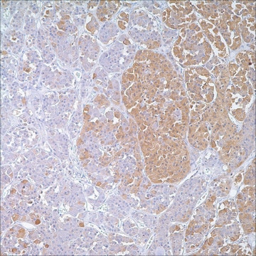
cytoplasmic

General description

Alpha-1-Antitrypsin is a protease inhibitor from the serpin superfamily, which inhibits a number of protease enzymes from inflammatory cells. It is an acute-phase plasma protein predominantly produced in hepatocytes, although it is also made in enterocytes and some mononuclear white blood cells. Alpha-1-antitrypsin immunohistochemistry is used to detect the accumulation of alpha-1-antitrypsin in hepatic lesions and histiocytic tumors.
The immunohistochemical staining of Alpha-1-Antitrypsin is considered to be very useful in the study of inherited AAT deficiency, benign and malignant hepatic tumors, and yolk sac carcinomas. Positive staining for A-1-Antitrypsin may also be used in the detection of benign and malignant lesions of a histiocytic nature. The sensitivity and specificity of the results have made this antibody a useful tool in the screening of patients with cryptogenic cirrhosis or other forms of liver disease with portal fibrosis of uncertain etiology.

Quality


IVD

IVD

IVD

RUO

Linkage

A-1-Antitrypsin Positive Control Slides , Product No. 223S, are available for immunohistochemistry (formalin-fixed, paraffin-embedded sections).

Physical form

Solution in Tris Buffer, pH 7.3-7.7, with 1% BSA and <0.1% Sodium Azide

Preparation Note

Download the IFU specific to your product lot and formatNote: This requires a keycode which can be found on your packaging or product label.

Other Notes

For Technical Service please contact: 800-665-7284 or email: service@cellmarque.com

Legal Information

Cell Marque is a trademark of Merck KGaA, Darmstadt, Germany

Not finding the right product?  

Try our Product Selector Tool.


Certificates of Analysis (COA)

Search for Certificates of Analysis (COA) by entering the products Lot/Batch Number. Lot and Batch Numbers can be found on a product’s label following the words ‘Lot’ or ‘Batch’.

Already Own This Product?

Find documentation for the products that you have recently purchased in the Document Library.

Visit the Document Library

H Takahashi et al.
Acta pathologica japonica, 40(9), 655-664 (1990-09-01)
Fifty-four adenoid cystic carcinomas (ACC) arising in major and minor salivary glands as well as in normal salivary glands were studied by immunohistochemistry for the presence of vimentin, neuron-specific enolase (NSE), alpha 1-antichymotrypsin (alpha 1-ACT) and alpha 1-antitrypsin (alpha 1-AT).
Alpha 1-antitrypsin in human macrophages.
P Isaacson et al.
Lancet (London, England), 2(8149), 964-965 (1979-11-03)
B Lindmark et al.
Histopathology, 16(3), 221-225 (1990-03-01)
We present a case of chronic liver disease with selective and exclusive hepatocyte endoplasmic reticulum storage of alpha 1-antichymotrypsin in the form of granules, detected by specific immunohistochemistry at the light microscopy level and corresponding to material found in dilated
L G Kindblom et al.
Human pathology, 13(9), 834-840 (1982-09-01)
The aim of this study was to localize alpha 1-antitrypsin, ferritin, and lysozyme by means of the indirect immunoperoxidase technique and to evaluate the significance of these antigens as markers of histiocytic differentiation in tumors of a supposed dual fibroblastic-histiocytic
Expression of protein markers in malignant hepatoma: evidence for genetic and epigenetic mechanisms.
P E Palmer et al.
Cancer, 45(6), 1424-1431 (1980-03-15)

Our team of scientists has experience in all areas of research including Life Science, Material Science, Chemical Synthesis, Chromatography, Analytical and many others.

Contact Technical Service