Skip to Content
Merck
All Photos(4)

Key Documents

HPA008729

Sigma-Aldrich

Anti-TMF1 antibody produced in rabbit

enhanced validation

Prestige Antibodies® Powered by Atlas Antibodies, affinity isolated antibody, buffered aqueous glycerol solution

Synonym(s):

Anti-TATA element modulatory factor antibody produced in rabbit, Anti-TMF antibody produced in rabbit

Sign Into View Organizational & Contract Pricing


About This Item

UNSPSC Code:
12352203
Human Protein Atlas Number:
NACRES:
NA.43

biological source

rabbit

Quality Level

conjugate

unconjugated

antibody form

affinity isolated antibody

antibody product type

primary antibodies

clone

polyclonal

product line

Prestige Antibodies® Powered by Atlas Antibodies

form

buffered aqueous glycerol solution

species reactivity

human, mouse, rat

enhanced validation

RNAi knockdown
Learn more about Antibody Enhanced Validation

technique(s)

immunoblotting: 0.04-0.4 μg/mL
immunofluorescence: 0.25-2 μg/mL
immunohistochemistry: 1:200-1:500

immunogen sequence

TSTTSDIEVLDHESVISESSASSRQETTDSKSSLHLMQTSFQLLSASACPEYNRLDDFQKLTESCCSSDAFERIDSFSVQSLDSRSVSEINSDDELSGKGYALVPIIVNSSTPKSKTVESAEGKSEEVNETLVIPTEEAEMEESG

UniProt accession no.

shipped in

wet ice

storage temp.

−20°C

target post-translational modification

unmodified

Gene Information

human ... TMF1(7110)

General description

TMF1 (TATA element modulatory factor 1) is a coiled-coil golgin protein, which is localized to the entire Golgi stack. This protein has a molecular weight of 123kDa, and is composed of 1093 amino acids. The corresponding gene is localized to human chromosome 3p12-p21. It contains at least three helical turns, and multiple putative leucin zippers. It is a Ser/Thr rich protein, with its N-terminal consisting of many serine-proline and threonine-proline repeats.

Immunogen

TATA element modulatory factor recombinant protein epitope signature tag (PrEST)

Application

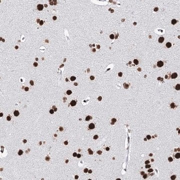
Anti-TMF1 antibody produced in rabbit, a Prestige Antibody, is developed and validated by the Human Protein Atlas (HPA) project . Each antibody is tested by immunohistochemistry against hundreds of normal and disease tissues. These images can be viewed on the Human Protein Atlas (HPA) site by clicking on the Image Gallery link. The antibodies are also tested using immunofluorescence and western blotting. To view these protocols and other useful information about Prestige Antibodies and the HPA, visit sigma.com/prestige.

Biochem/physiol Actions

TMF1 (TATA element modulatory factor 1) is suggested to have trans-Golgi functions. It is a putative transcription factor, which regulates the function of cellular as well as viral genes. In gel-retardation assays, it interacts with and binds to the TATA element of HIV1 (human immunodeficiency virus) and prevents its activation. Its genetic locus is subjected to frequent rearrangements in renal and lung carcinomas. During cellular stress conditions, TMF1 gets activated and changes its subcellular localization and activates multiple proteins, including Stat3, a transcription factor. Stat3 then undergoes proteasomal degradation. Its expression is suppressed in solid tumors. As it controls multiple transcription factors during cellular stress conditions, it influences angiogenesis and progression of solid tumors, such as prostate cancer.

Features and Benefits

Prestige Antibodies® are highly characterized and extensively validated antibodies with the added benefit of all available characterization data for each target being accessible via the Human Protein Atlas portal linked just below the product name at the top of this page. The uniqueness and low cross-reactivity of the Prestige Antibodies® to other proteins are due to a thorough selection of antigen regions, affinity purification, and stringent selection. Prestige antigen controls are available for every corresponding Prestige Antibody and can be found in the linkage section.

Every Prestige Antibody is tested in the following ways:
  • IHC tissue array of 44 normal human tissues and 20 of the most common cancer type tissues.
  • Protein array of 364 human recombinant protein fragments.

Linkage

Corresponding Antigen APREST70930

Physical form

Solution in phosphate-buffered saline, pH 7.2, containing 40% glycerol and 0.02% sodium azide

Legal Information

Prestige Antibodies is a registered trademark of Merck KGaA, Darmstadt, Germany

Disclaimer

Unless otherwise stated in our catalog or other company documentation accompanying the product(s), our products are intended for research use only and are not to be used for any other purpose, which includes but is not limited to, unauthorized commercial uses, in vitro diagnostic uses, ex vivo or in vivo therapeutic uses or any type of consumption or application to humans or animals.

Not finding the right product?  

Try our Product Selector Tool.

Storage Class Code

10 - Combustible liquids

WGK

WGK 1

Flash Point(F)

Not applicable

Flash Point(C)

Not applicable

Personal Protective Equipment

dust mask type N95 (US), Eyeshields, Gloves

Certificates of Analysis (COA)

Search for Certificates of Analysis (COA) by entering the products Lot/Batch Number. Lot and Batch Numbers can be found on a product’s label following the words ‘Lot’ or ‘Batch’.

Already Own This Product?

Find documentation for the products that you have recently purchased in the Document Library.

Visit the Document Library

Shai Bel et al.
The Journal of biological chemistry, 287(30), 25631-25639 (2012-05-04)
TMF/ARA160 is a Golgi-associated protein with several cellular functions, among them direction of the NF-κB subunit, p65 RelA, to ubiquitination and proteasomal degradation in stressed cells. We sought to investigate the role of TMF/ARA160 under imposed stress conditions in vivo.
Galya Abrham et al.
International journal of cancer, 125(1), 43-53 (2009-03-31)
TMF/ARA160 is a Golgi-associated protein whose level is downregulated in solid tumors. TMF changes its subcellular localization on exposure of cells to stress cues, thereby, directing proteins, such as the key transcription factor, Stat3, to proteasomal degradation. Here, we show
Malin Lando et al.
The Journal of pathology, 230(1), 59-69 (2013-01-22)
The pathogenetic role, including its target genes, of the recurrent 3p12-p14 loss in cervical cancer has remained unclear. To determine the onset of the event during carcinogenesis, we used microarray techniques and found that the loss was the most frequent
Akihiro Harada et al.
Nature communications, 15(1), 4514-4514 (2024-05-28)
Knowledge on the distribution and dynamics of glycosylation enzymes in the Golgi is essential for better understanding this modification. Here, using a combination of CRISPR/Cas9 knockin technology and super-resolution microscopy, we show that the Golgi complex is assembled by a
Victoria J Miller et al.
The Journal of biological chemistry, 288(6), 4229-4240 (2012-12-15)
Protein sorting between eukaryotic compartments requires vesicular transport, wherein tethering provides the first contact between vesicle and target membranes. Here we map and start to functionally analyze the interaction network of the conserved oligomeric Golgi (COG) complex that mediates retrograde

Our team of scientists has experience in all areas of research including Life Science, Material Science, Chemical Synthesis, Chromatography, Analytical and many others.

Contact Technical Service