Skip to Content
Merck
All Photos(1)

Key Documents

I1784

Sigma-Aldrich

Anti-Importin α antibody, Mouse monoclonal

clone IM-75, purified from hybridoma cell culture

Synonym(s):

Anti-Imp α, Anti-Qip1, Anti-karyopherin α

Sign Into View Organizational & Contract Pricing


About This Item

MDL number:
UNSPSC Code:
12352203
NACRES:
NA.41

biological source

mouse

conjugate

unconjugated

antibody form

purified immunoglobulin

antibody product type

primary antibodies

clone

IM-75, monoclonal

form

buffered aqueous solution

mol wt

antigen ~60 kDa

species reactivity

bovine, human, mouse, rat

technique(s)

immunocytochemistry: suitable
immunoprecipitation (IP): suitable
indirect ELISA: suitable
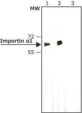
western blot: 1-2 μg/mL using HeLa cell extract

isotype

IgG2b

UniProt accession no.

shipped in

dry ice

storage temp.

−20°C

target post-translational modification

unmodified

Gene Information

human ... KPNA4(3840)
mouse ... Kpna4(16649)
rat ... Kpna4(361959)

General description

Monoclonal Anti-Importin α (mouse IgG2b isotype) is derived from the hybridoma IM-75 produced by the fusion of mouse myeloma cells (NS1 cells), and splenocytes from BALB/c mice, immunized with recombinant human importin α. The Importin α family of proteins is comprised of three subfamilies based on amino acid sequence similarity: SRP1-like subfamily (containing the SRP1, importin α5, α6 and α7), Rch1-like subfamily (containing Rch1, pendulin, importin α2), and α3/Qip1-like subfamily (containing α4).

Immunogen

recombinant human importin α.

Application

Anti-Importin α antibody, Mouse monoclonal has been used:
  • enzyme linked immunosorbent assay (ELISA)
  • immunoblotting
  • immunoprecipitation
  • immunocytochemistry

Monoclonal Anti-Importin α antibody produced in mouse has been used in western blotting.

Biochem/physiol Actions

The gene KPNA4 (karyopherin subunit α 4) encodes a 16kDa protein, also referred to as importin α 3 belonging to the family of karyopherins (designated KPNA1 to KPNA4). It participates in the recognition of NLS (nuclear localization sequence) sites and dock NLS-containing proteins destined for transport into the nuclear pore complex. It binds to translocon and participates in the sorting of INM (inner nuclear membrane)-directed integral membrane proteins. It associates with nucleoporins and functions in cargo-translocation across the nuclear membrane. Each KPNA contains an N-terminal repeat domain called the importin β binding site that binds to KPNB1, which in turn directs each KPNA/cargo complex through the nuclear pore. The encoded shuttling protein also functions in NF (nuclear factor)-κB nuclear translocation.

Physical form

Solution in 0.01 M phosphate buffered saline, pH 7.4, and 15 mM sodium azide.

Disclaimer

Unless otherwise stated in our catalog or other company documentation accompanying the product(s), our products are intended for research use only and are not to be used for any other purpose, which includes but is not limited to, unauthorized commercial uses, in vitro diagnostic uses, ex vivo or in vivo therapeutic uses or any type of consumption or application to humans or animals.

Not finding the right product?  

Try our Product Selector Tool.

Storage Class Code

10 - Combustible liquids

WGK

nwg

Flash Point(F)

Not applicable

Flash Point(C)

Not applicable


Certificates of Analysis (COA)

Search for Certificates of Analysis (COA) by entering the products Lot/Batch Number. Lot and Batch Numbers can be found on a product’s label following the words ‘Lot’ or ‘Batch’.

Already Own This Product?

Find documentation for the products that you have recently purchased in the Document Library.

Visit the Document Library

Jin Xu et al.
Oncology reports, 30(4), 1878-1882 (2013-08-01)
Recent studies have shown the localization of RhoA in the cell nucleus, in addition to its cellular distribution in the cytosol and cell membrane. Our previous results that a high amount of RhoA was detected in gastric cancer cell nucleus
Laura Padron-Barthe et al.
Molecular and cellular biology, 27(11), 4028-4036 (2007-04-04)
The best-characterized biochemical feature of apoptosis is degradation of genomic DNA into oligonucleosomes. The endonuclease responsible for DNA degradation in caspase-dependent apoptosis is caspase-activated DNase. In caspase-independent apoptosis, different endonucleases may be activated according to the cell line and the
Guillaume Huet et al.
Journal of cell science, 126(Pt 2), 497-507 (2012-12-04)
Phactr proteins bind actin and protein phosphatase 1 (PP1), and are involved in processes ranging from angiogenesis to cell cycle regulation. Phactrs share a highly conserved RPEL domain with the myocardin-related transcription factor (MRTF) family, where actin binding to this
KPNA4 Mediates Vitamin D-Dependent Inhibition of NF-kB Activity in Swine Epicardial Preadipocytes.
Chen, et al.
Circulation, 130, A12818-A12818 (2014)
Aging impairs transcriptional regulation of vascular endothelial growth factor in human microvascular endothelial cells: implications for angiogenesis and cell survival.
Ahluwalia, A., et al.
Journal of Physiology And Pharmacology, 65 (2014)

Our team of scientists has experience in all areas of research including Life Science, Material Science, Chemical Synthesis, Chromatography, Analytical and many others.

Contact Technical Service