Skip to Content
Merck
All Photos(4)

Key Documents

SAB2700248

Sigma-Aldrich

Anti-POM121 antibody produced in rabbit

affinity isolated antibody, buffered aqueous solution

Synonym(s):

DKFZp586G1822, DKFZp586P2220, FLJ41820, POM121

Sign Into View Organizational & Contract Pricing


About This Item

UNSPSC Code:
12352203
NACRES:
NA.43

Pricing and availability is not currently available.

biological source

rabbit

Quality Level

conjugate

unconjugated

antibody form

affinity isolated antibody

antibody product type

primary antibodies

clone

polyclonal

form

buffered aqueous solution

species reactivity

human, mouse

technique(s)

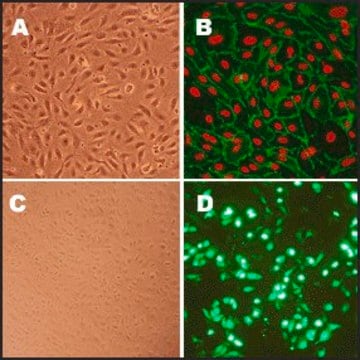
immunofluorescence: suitable
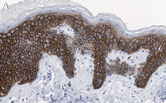
immunohistochemistry: suitable
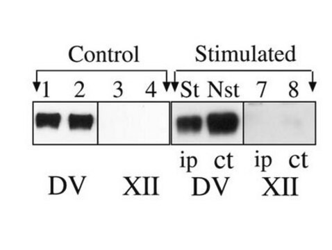
western blot: 500-3000

NCBI accession no.

Immunogen

Recombinant fragment corresponding to a region within amino acids 236 and 562 of POM121 according to NP_742017

Application

Suggested starting dilutions are as follows: ICC/IF: 1:100-1:1000, IHC-P: 1:100-1:1000, WB: 1:500-1:3000. Not yet tested in other applications. Optimal working dilutions should be determined experimentally by the end user.

Biochem/physiol Actions

The nuclear envelope creates distinct nuclear and cytoplasmic compartments in eukaryotic cells. It consists of two concentric membranes perforated by nuclear pores, large protein complexes that form aqueous channels to regulate the flow of macromolecules between the nucleus and the cytoplasm. These complexes are composed of at least 100 different polypeptide subunits, many of which belong to the nucleoporin family. This gene encodes a member of the FG-repeat-containing nucleoporins. The protein encoded by this gene is an integral membrane protein that localizes to the central spoke ring complex and participates in anchoring the nuclear pore complex to the nuclear envelope. Alternatively spliced variants that encode different protein isoforms have been described but the full-length nature of only one has been determined. [provided by RefSeq]

Features and Benefits

Evaluate our antibodies with complete peace of mind. If the antibody does not perform in your application, we will issue a full credit or replacement antibody. Learn more.

Physical form

1XPBS, 1% BSA, 20% Glycerol (pH7). 0.025% ProClin 300 was added as a preservative.

Disclaimer

Unless otherwise stated in our catalog or other company documentation accompanying the product(s), our products are intended for research use only and are not to be used for any other purpose, which includes but is not limited to, unauthorized commercial uses, in vitro diagnostic uses, ex vivo or in vivo therapeutic uses or any type of consumption or application to humans or animals.

Not finding the right product?  

Try our Product Selector Tool.

Pictograms

Exclamation mark

Signal Word

Warning

Hazard Statements

Hazard Classifications

Aquatic Chronic 3 - Skin Sens. 1

Storage Class Code

12 - Non Combustible Liquids

WGK

WGK 2

Flash Point(F)

Not applicable

Flash Point(C)

Not applicable


Certificates of Analysis (COA)

Search for Certificates of Analysis (COA) by entering the products Lot/Batch Number. Lot and Batch Numbers can be found on a product’s label following the words ‘Lot’ or ‘Batch’.

Need A Sample COA?

This is a sample Certificate of Analysis (COA) and may not represent a recently manufactured lot of this specific product.

Already Own This Product?

Find documentation for the products that you have recently purchased in the Document Library.

Visit the Document Library

Jennifer M Röhrl et al.
Cells, 10(1) (2021-01-21)
Hutchinson-Gilford progeria syndrome (HGPS) is a rare premature aging disease caused by a mutation in LMNA. A G608G mutation in exon 11 of LMNA is responsible for most HGPS cases, generating a truncated protein called "progerin". Progerin is permanently farnesylated
Debora Keller et al.
iScience, 27(4), 109343-109343 (2024-03-21)
Spatial genome organization within the nucleus influences major biological processes and is impacted by the configuration of linear chromosomes. Here, we applied 3D spatial statistics and modeling on high-resolution telomere and centromere 3D-structured illumination microscopy images in cancer cells. We

Our team of scientists has experience in all areas of research including Life Science, Material Science, Chemical Synthesis, Chromatography, Analytical and many others.

Contact Technical Service