Skip to Content
Merck
All Photos(8)

Key Documents

HPA008346

Sigma-Aldrich

ANTI-SUN1 antibody produced in rabbit

enhanced validation

Ab1, Prestige Antibodies® Powered by Atlas Antibodies, affinity isolated antibody, buffered aqueous glycerol solution

Synonym(s):

Anti-Sad1/unc-84 protein-like 1, Anti-UNC84A, Anti-Unc-84 homolog A

Sign Into View Organizational & Contract Pricing


About This Item

UNSPSC Code:
12352203
Human Protein Atlas Number:
NACRES:
NA.43

biological source

rabbit

Quality Level

conjugate

unconjugated

antibody form

affinity isolated antibody

antibody product type

primary antibodies

clone

polyclonal

product line

Prestige Antibodies® Powered by Atlas Antibodies

form

buffered aqueous glycerol solution

species reactivity

human

enhanced validation

orthogonal RNAseq
independent
Learn more about Antibody Enhanced Validation

technique(s)

immunofluorescence: 0.25-2 μg/mL
immunohistochemistry: 1:200-1:500

immunogen sequence

QLLPTVEHLQLELDQLKSELSSWRHVKTGCETVDAVQERVDVQVREMVKLLFSEDQQGGSLEQLLQRFSSQFVSKGDLQTMLRDLQLQILRNVTHHVSVTKQLPTSEAVVSAVSEAGASGITEAQARAIVNS

shipped in

wet ice

storage temp.

−20°C

target post-translational modification

unmodified

Gene Information

human ... UNC84A(23353)

Looking for similar products? Visit Product Comparison Guide

General description

Sad1 and UNC84 domain containing 1 (SUN1) is present in the inner nuclear envelope. It has a carboxyl-terminal domain called the SUN domain which is located in the space between the inner and outer nuclear membranes and an amino-terminal domain located in the nucleoplasm.
The SUN1 gene is localized on human chromosome 7p22.3. It is ubiquitously expressed and exists as 10 variants due to alternative splicing events.

Immunogen

Sad1/unc-84 protein-like 1 recombinant protein epitope signature tag (PrEST)

Application

Anti-SUN1 antibody produced in rabbit has been used in immunoprecipitation.
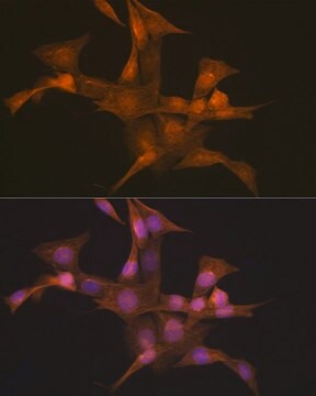
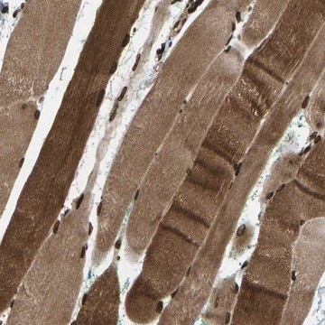
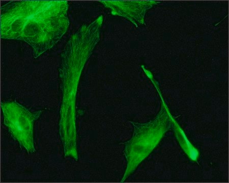
Anti-SUN1 antibody produced in rabbit, a Prestige Antibody, is developed and validated by the Human Protein Atlas (HPA) project . Each antibody is tested by immunohistochemistry against hundreds of normal and disease tissues. These images can be viewed on the Human Protein Atlas (HPA) site by clicking on the Image Gallery link. The antibodies are also tested using immunofluorescence and western blotting. To view these protocols and other useful information about Prestige Antibodies and the HPA, visit sigma.com/prestige.
Applications in which this antibody has been used successfully, and the associated peer-reviewed papers, are given below.
Immunocytochemistry (1 paper)
Immunohistochemistry (1 paper)
Western Blotting (1 paper)
These images and the collection of this vast data set can be viewed on the Human Protein Atlas (HPA) site by clicking on the Image Gallery link. We also provide Prestige Antibodies® protocols and other useful information.

Biochem/physiol Actions

Sad1 and UNC84 domain containing 1 (SUN1) plays an important role in linking telomeres with the nuclear envelope. It binds to telomere-binding proteins and helps in the formation of the meiotic bouquet. This is a special cluster formed by telomeres at the nuclear envelope and occurs before meiosis I. SUN1 also interacts with SUN2 to form heterodimers. This in turn helps in aiding the interaction between the nuclear envelope and the centrosome. It has also been shown that reforming nuclear envelope employs SUN1 to acetylate histones for de-condensation of DNA after mitosis. It is the one of the earliest factors to associate with segregated daughter chromosomes in anaphase.

Features and Benefits

Prestige Antibodies® are highly characterized and extensively validated antibodies with the added benefit of all available characterization data for each target being accessible via the Human Protein Atlas portal linked just below the product name at the top of this page. The uniqueness and low cross-reactivity of the Prestige Antibodies® to other proteins are due to a thorough selection of antigen regions, affinity purification, and stringent selection. Prestige antigen controls are available for every corresponding Prestige Antibody and can be found in the linkage section.

Every Prestige Antibody is tested in the following ways:
  • IHC tissue array of 44 normal human tissues and 20 of the most common cancer type tissues.
  • Protein array of 364 human recombinant protein fragments.

Linkage

Corresponding Antigen APREST71108

Physical form

Solution in phosphate-buffered saline, pH 7.2, containing 40% glycerol and 0.02% sodium azide

Legal Information

Prestige Antibodies is a registered trademark of Merck KGaA, Darmstadt, Germany

Disclaimer

Unless otherwise stated in our catalog or other company documentation accompanying the product(s), our products are intended for research use only and are not to be used for any other purpose, which includes but is not limited to, unauthorized commercial uses, in vitro diagnostic uses, ex vivo or in vivo therapeutic uses or any type of consumption or application to humans or animals.

Not finding the right product?  

Try our Product Selector Tool.

Storage Class Code

10 - Combustible liquids

WGK

WGK 1

Personal Protective Equipment

dust mask type N95 (US), Eyeshields, Gloves

Certificates of Analysis (COA)

Search for Certificates of Analysis (COA) by entering the products Lot/Batch Number. Lot and Batch Numbers can be found on a product’s label following the words ‘Lot’ or ‘Batch’.

Already Own This Product?

Find documentation for the products that you have recently purchased in the Document Library.

Visit the Document Library

Monika Zwerger et al.
Molecular biology of the cell, 21(2), 354-368 (2009-11-27)
Lamin B receptor (LBR) is an inner nuclear membrane protein involved in tethering the nuclear lamina and the underlying chromatin to the nuclear envelope. In addition, LBR exhibits sterol reductase activity. Mutations in the LBR gene cause two different human
Stefan Hintze et al.
Frontiers in physiology, 9, 1532-1532 (2018-11-15)
Myotonic dystrophy type 1 (DM1) is a multisystemic disorder with predominant myotonia and muscular dystrophy which is caused by CTG-repeat expansions in the DMPK gene. These repeat expansions are transcribed and the resulting mRNA accumulates RNA-binding proteins involved in splicing
Xiang Lu et al.
Cells, 7(4) (2018-04-25)
The mammalian nuclear lamina proteins—prelamin A- and B-type lamins—are post-translationally modified by farnesylation, endoproteolysis, and carboxymethylation at a carboxy-terminal CAAX (C, cysteine; a, aliphatic amino acid; X, any amino acid) motif. However, prelamin A processing into mature lamin A is
Laure Crabbe et al.
Cell reports, 2(6), 1521-1529 (2012-12-25)
Telomeres are essential for nuclear organization in yeast and during meiosis in mice. Exploring telomere dynamics in living human cells by advanced time-lapse confocal microscopy allowed us to evaluate the spatial distribution of telomeres within the nuclear volume. We discovered
Ya-Hui Chi et al.
The Journal of biological chemistry, 282(37), 27447-27458 (2007-07-17)
Replicated mammalian chromosomes condense to segregate during anaphase, and they de-condense at the conclusion of mitosis. Currently, it is not understood what the factors and events are that specify de-condensation. Here, we demonstrate that chromosome de-condensation needs the function of

Our team of scientists has experience in all areas of research including Life Science, Material Science, Chemical Synthesis, Chromatography, Analytical and many others.

Contact Technical Service