Skip to Content
Merck
All Photos(1)

Documents

C1956

Sigma-Aldrich

Monoclonal Anti-Calcineurin (α-Subunit) antibody produced in mouse

clone CN-A1, ascites fluid

Sign Into View Organizational & Contract Pricing


About This Item

MDL number:
UNSPSC Code:
12352203
NACRES:
NA.44

biological source

mouse

Quality Level

conjugate

unconjugated

antibody form

ascites fluid

antibody product type

primary antibodies

clone

CN-A1, monoclonal

contains

15 mM sodium azide

species reactivity

human, bovine, rat

technique(s)

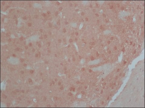
immunohistochemistry (formalin-fixed, paraffin-embedded sections): suitable using neurons in human ganglia
indirect ELISA: suitable
microarray: suitable
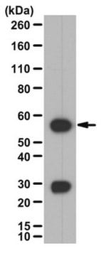
western blot: 1:10,000 using rat brain extract

isotype

IgG1

UniProt accession no.

shipped in

dry ice

storage temp.

−20°C

target post-translational modification

unmodified

Gene Information

human ... PPP3R1(5534)
rat ... Ppp3r1(29748)

General description

Calcineurin (CaN)(heterodimeric enzyme) is a Ca2+/calmodulin (CaM)-stimulated protein phosphatase 3 (PPP3) consisting of a catalytic A subunit (CnA) and a Ca2+-binding regulatory B subunit (CnB). The calcium-binding regulatory subunit (calcineurin B) is coded by the PPP3R1 gene located on human chromosome 2. Protein phosphatase 3, regulatory subunit B, α (PPP3R1) is also known as CALN, CCN1, CNA1, CALNA, PPP2B and CALNA1. The regulatory subunit calcineurin B is widely expressed in brain while the catalytic subunit (calcineurin A) is abundantly expressed in Hassall′s corpuscles, localized in the thymic medulla and represents the terminal stages of thymic medullary epithelium.
Monoclonal Anti-Calcineurin (α-subunit) (mouse IgG1 isotype) is derived from the CN-A1 hybridoma produced by the fusion of mouse myeloma cells and splenocytes from immunized BALB/c mice.

Specificity

In immunoblotting, the product recognizes an epitope located on the α-subunit of calcineurin (61 kDa, also called calcineurin A) and does not cross-react with the β-subunit.

Immunogen

bovine brain calcineurin

Application

Monoclonal Anti-Calcineurin (α-Subunit) antibody produced in mouse has been used in:enzyme linked immuno sorbent assay (ELISA) ,immunohistochemistry, western blotting

Biochem/physiol Actions

Calcineurin is a major soluble calmodulin-binding protein in the brain. It stimulates the expression of the surface molecules CD83, CD80, CD86, CD40 and HLA-DR by promoting secretion of inflammatory cytokines IL-6, TNF-α and IL-1β by human PBMC-derived dendritic cells. It plays an important role in signal transduction, activation of T cell. Calcineurin is an excellent marker enzyme for the detection of neuronal activity and synaptic plasticity after brain damage.

Disclaimer

Unless otherwise stated in our catalog or other company documentation accompanying the product(s), our products are intended for research use only and are not to be used for any other purpose, which includes but is not limited to, unauthorized commercial uses, in vitro diagnostic uses, ex vivo or in vivo therapeutic uses or any type of consumption or application to humans or animals.

Not finding the right product?  

Try our Product Selector Tool.

Storage Class Code

10 - Combustible liquids

WGK

nwg

Flash Point(F)

Not applicable

Flash Point(C)

Not applicable


Certificates of Analysis (COA)

Search for Certificates of Analysis (COA) by entering the products Lot/Batch Number. Lot and Batch Numbers can be found on a product’s label following the words ‘Lot’ or ‘Batch’.

Already Own This Product?

Find documentation for the products that you have recently purchased in the Document Library.

Visit the Document Library

H Bito et al.
Cell, 87(7), 1203-1214 (1996-12-27)
While changes in gene expression are critical for many brain functions, including long-term memory, little is known about the cellular processes that mediate stimulus-transcription coupling at central synapses. In studying the signaling pathways by which synaptic inputs control the phosphorylation
Colin Rickman et al.
The Journal of biological chemistry, 279(13), 12574-12579 (2004-01-08)
Synaptotagmins are membrane proteins that possess tandem C2 domains and play an important role in regulated membrane fusion in metazoan organisms. Here we show that both synaptotagmins I and II, the two major neuronal isoforms, can interact with the syntaxin/synaptosomal-associated
Yasuko Hirakawa et al.
Journal of molecular signaling, 4, 6-6 (2009-09-04)
Glucocorticoid hormones (GCs) induce apoptosis of leukemic T-cells by transcriptional regulation via the GC receptor, GR. In the human leukemic CEM cell culture model, RCAN1 has been identified as one of the genes that is specifically upregulated only in the
Y Oishi et al.
Acta physiologica (Oxford, England), 192(3), 381-395 (2007-09-26)
We used a model of chronic unloading followed by reloading to examine the apoptotic responses associated with soleus muscle atrophy and subsequent recovery. Male Wistar rats were subjected to hindlimb unloading (HU) for 2 weeks and subsequent reloading for 0
Maya Fujita et al.
Biological & pharmaceutical bulletin, 41(5), 786-796 (2018-05-02)
Although calcineurin is abundantly expressed in the nervous system and involved in neurite extension and synaptic plasticity in neurons, little is known about its roles in glial cells. To investigate the roles of calcineurin in glial cells, we generated glial

Our team of scientists has experience in all areas of research including Life Science, Material Science, Chemical Synthesis, Chromatography, Analytical and many others.

Contact Technical Service