immunohistochemistry (formalin-fixed, paraffin-embedded sections): 1:500 using sections of rat, human or chicken cerebellum sections microarray: suitable western blot: 1:1,000 using recombinant rat calbindin-D-28K
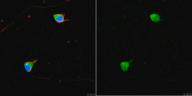
Calbindin-D-28K (also termed vitamin D-dependent calcium-binding protein, or cholecalcin), is a highly conserved 28 kDa calcium binding protein, with broad tissue distribution. Itbelongs , to a family of low molecular weight calcium-binding proteins (CaBPs). Calbindin-D-28K is found predominantly in subpopulations of central and peripheral nervous system neurons, and in certain epithelial cells.
Immunogen
synthetic peptide corresponding to the C-terminal region of rat calbindin-D-28K (amino acids 185-199). This sequence is identical in the corresponding human, mouse and bovine calbindin-D-28K sequences and is highly conserved (single amino acid substitution) in chicken and frog calbindin-D-28K.
Application
Anti-Calbindin-D-28K (KD-15) antibody produced in rabbit has been used in:
Calbindin 1 (CALB) is a calcium-binding protein. The protein may play a role in development of Purkinje cells found in heterotopias and cerebellar dysgenesias. It also acts as a very important component of intracellular homeostasis in cerebellar neurons. The progressive decrease of calbindin content in the Purkinje cells undergoes degeneration and death during paraneoplastic changes in the cerebellum. Calbindin effects on Parkinson′s disease (PD) risk displays population specificity.
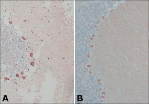
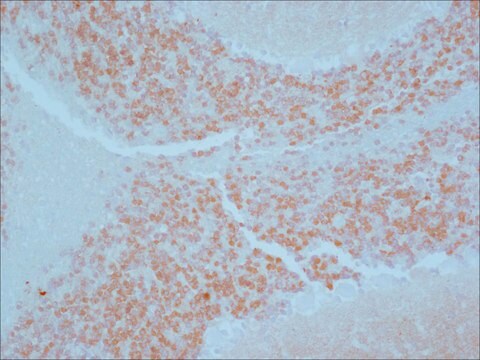
Calbindin-D-28K is an important neuroanatomical marker. Despite its broad tissue distribution, it exhibits a cell-type-specific expression pattern. It has been immunocytochemically localized in selected cells in hippocampus, where it is thought to buffer intracellular calcium, or transport intramembranous calcium.
Physical form
Solution in 0.01 M phosphate buffered saline, pH 7.4, containing 1% bovine serum albumin and 15 mM sodium azide.
Analysis Note
Enzymatic predigestion of formalin-fixed, paraffin-embedded sections by proteolytic enzymes (e.g., 0.1% trypsin or protease, 10 min. at RT or 37 °C) improves immunohistochemical staining.
Disclaimer
Unless otherwise stated in our catalog or other company documentation accompanying the product(s), our products are intended for research use only and are not to be used for any other purpose, which includes but is not limited to, unauthorized commercial uses, in vitro diagnostic uses, ex vivo or in vivo therapeutic uses or any type of consumption or application to humans or animals.
The Journal of comparative neurology, 501(4), 619-630 (2007-02-06)
Purkinje cells in the cerebellum express the antigen zebrin II (aldolase C) in many vertebrates. In mammals, zebrin is expressed in a parasagittal fashion, with alternating immunopositive and immunonegative stripes. Whether a similar pattern is expressed in birds is unknown.
Subpopulations of neurons expressing parvalbumin in the human amygdala
Pantazopoulos H, et al.
The Journal of Comparative Neurology, 496(5), 706-722 (2006)
Distribution of and steroid hormone effects on calbindin-D9k in the immature rat brain
Park SY, et al.
Brain Research Bulletin, 152, 225-235 (2019)
Altered hippocampal expression of calbindin-D-28k and calretinin in GABAB (1)-deficient mice
Our team of scientists has experience in all areas of research including Life Science, Material Science, Chemical Synthesis, Chromatography, Analytical and many others.