Skip to Content
Merck
All Photos(2)

Documents

05-829-AF647

Sigma-Aldrich

Anti-α-Tubulin Antibody, clone DM1A, Alexa Fluor 647 conjugate

clone DM1A, from mouse, ALEXA FLUOR 647

Synonym(s):

Tubulin alpha-1 chain, α-Tubulin

Sign Into View Organizational & Contract Pricing


About This Item

UNSPSC Code:
12352203
eCl@ss:
32160702
NACRES:
NA.41

biological source

mouse

Quality Level

conjugate

ALEXA FLUOR 647

antibody form

purified immunoglobulin

antibody product type

primary antibodies

clone

DM1A, monoclonal

species reactivity

guinea pig, rat, porcine, human, gerbil, mouse, bovine, avian

technique(s)

immunocytochemistry: suitable

isotype

IgG1

NCBI accession no.

UniProt accession no.

shipped in

wet ice

target post-translational modification

unmodified

Gene Information

human ... TUBA1A(7846)

General description

Tubulin alpha-1 chain (UniProt P02552) is encoded by the TUBA1C (also known as TUBA1) gene (Gene ID 429035) in Gallus gallus (Chicken) species. Microtubules are cytoskeletal filaments composed of alpha-/beta-tubulin heterodimers that self-assemble head-to-tail to form protofilaments and laterally to form a hollow tube. Various post-translational modifications, including acetylation, polyglutamylation, polyglycylation, detyrosination, phosphorylation, and palmitoylation, can regulate the polymerization properties of tubulins and/or their interactions with microtubule associated proteins (MAPs) and motor proteins.

Specificity

Clone DM1A recognizes α-Tubulin, MW 50-60 kDa. The epitope recognized by clone DM1A has been mapped to the C-terminal 25 amino acids.

Immunogen

Epitope: C-terminal 25 amino acids.
Purified chicken brain α-Tubulin. The epitope recognized by this antibody has been mapped to the C-terminal 25 amino acids (AALEKDYEEVGVDSVEGEGEEEGEE).

Application

Anti-α-Tubulin Antibody, clone DM1A, Alexa Fluor 647 conjugate is an antibody against α-Tubulin for use in Immunocytochemistry.
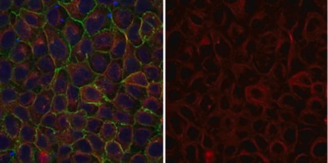
Immunocytochemistry Analysis: A 1:100 dilution from a representative lot detected α-Tubulin in NIH/3T3 cells.
The unconjugated antibody (Cat. No. 05-829) is shown to be suitable also for immunocytochemistry, immunofluorescence, and Western blotting applications.
Research Category
Cell Structure
Research Sub Category
Cytoskeleton

Quality

Evaluated by Immunocytochemistry in A431 cells.

Immunocytochemistry Analysis: A 1:100 dilution of this antibody detected α-Tubulin in A431 cells.

Target description

~50 kDa observed.

Physical form

Protein G Purified
Purified mouse monoclonal IgG1 antibody conjugate in PBS with 15 mg/mL BSA and 0.1 % sodium azide.

Storage and Stability

Stable for 1 year at 2-8°C from date of receipt.

Other Notes

Concentration: Please refer to lot specific datasheet.

Legal Information

ALEXA FLUOR is a trademark of Life Technologies

Disclaimer

Unless otherwise stated in our catalog or other company documentation accompanying the product(s), our products are intended for research use only and are not to be used for any other purpose, which includes but is not limited to, unauthorized commercial uses, in vitro diagnostic uses, ex vivo or in vivo therapeutic uses or any type of consumption or application to humans or animals.

Not finding the right product?  

Try our Product Selector Tool.

Storage Class Code

12 - Non Combustible Liquids

WGK

WGK 2

Flash Point(F)

Not applicable

Flash Point(C)

Not applicable


Certificates of Analysis (COA)

Search for Certificates of Analysis (COA) by entering the products Lot/Batch Number. Lot and Batch Numbers can be found on a product’s label following the words ‘Lot’ or ‘Batch’.

Already Own This Product?

Find documentation for the products that you have recently purchased in the Document Library.

Visit the Document Library

Brandon Weissbourd et al.
Cell, 184(24), 5854-5868 (2021-11-26)
Jellyfish are radially symmetric organisms without a brain that arose more than 500 million years ago. They achieve organismal behaviors through coordinated interactions between autonomously functioning body parts. Jellyfish neurons have been studied electrophysiologically, but not at the systems level.
David J Schodt et al.
Biomedical optics express, 14(1), 429-440 (2023-01-27)
We describe a dedicated microscope for automated sequential localization microscopy which we term Sequential Super-resolution Microscope (SeqSRM). This microscope automates precise stage stabilization on the order of 5-10 nanometers and data acquisition of all user-selected cells on a coverslip, limiting
Farah Haque et al.
Nature cell biology, 24(7), 1088-1098 (2022-06-21)
A long-established strategy for transcription regulation is the tethering of transcription factors to cellular membranes. By contrast, the principal effectors of Hedgehog signalling, the GLI transcription factors, are regulated by microtubules in the primary cilium and the cytoplasm. How GLI
Luqman O Awoniyi et al.
Journal of cell science, 136(15) (2023-07-07)
Successful B cell activation, which is critical for high-affinity antibody production, is controlled by the B cell antigen receptor (BCR). However, we still lack a comprehensive protein-level view of the very dynamic multi-branched cellular events triggered by antigen binding. Here

Our team of scientists has experience in all areas of research including Life Science, Material Science, Chemical Synthesis, Chromatography, Analytical and many others.

Contact Technical Service