Skip to Content
Merck
All Photos(2)

Key Documents

MAB3234

Sigma-Aldrich

Anti-Cytokeratin 18 Antibody, clone RGE53

clone RGE53, Chemicon®, from mouse

Sign Into View Organizational & Contract Pricing


About This Item

UNSPSC Code:
12352203
eCl@ss:
32160702
NACRES:
NA.41

biological source

mouse

Quality Level

antibody form

purified immunoglobulin

antibody product type

primary antibodies

clone

RGE53, monoclonal

species reactivity

human, pig, mouse, chicken, canine, rat, rabbit, hamster

manufacturer/tradename

Chemicon®

technique(s)

flow cytometry: suitable
immunocytochemistry: suitable
immunohistochemistry: suitable
western blot: suitable

isotype

IgG1

suitability

not suitable for immunohistochemistry (Paraffin)

NCBI accession no.

UniProt accession no.

shipped in

dry ice

target post-translational modification

unmodified

Gene Information

human ... KRT18(3875)

General description

Cytokeratins are a subfamily of intermediate filament proteins and are characterized by a remarkable biochemical diversity, represented in epithelial tissues by at least 20 different polypeptides. They range in molecular weight from between 40 kDa and 68 kDa and isoelectric pH between 4.9 - 7.8, The individual cytokeratin polypeptides are designated 1-20. The various epithelia in the human body usually express cytokeratins which are not only characteristic of the type of epithelium, but also related to the degree of maturation or differentiation within an epithelium. Cytokeratin subtype expression patterns are used to an increasing extent in the distinction of different types of epithelial malignancies.

Specificity

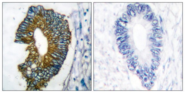
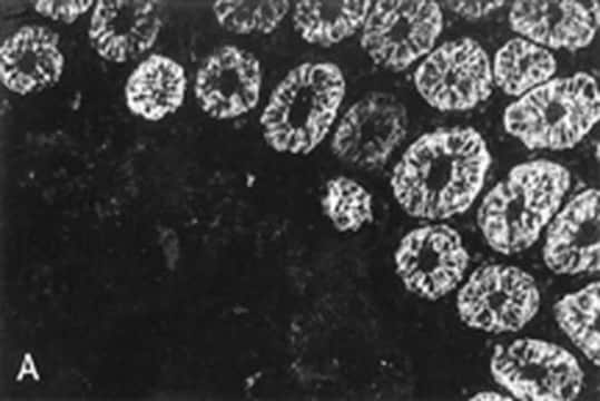
Specifically recognizes glandular epithelial cells from gastro-intestinal tract, respiratory tract and urogenetical tract. As well as endocrine and exocrine tissues and mesothelial cells. It is unreactive with stratified squamous epithelia (esophagus, epidermis, ectocervix). In transitional epithelium of the bladder RGE53 only reacts with the superficial (umbrella) cells. RGE53 can distinguish adenocarcinomas and mesotheliomas from squamous cell carcinoma and non-epithelial tumours. The antibody is directed against 45 kD cytokeratin protein (cytokeratin 18). Cross reactivity with; Keratin from most mammals, birds.

Immunogen

Cytoskeletal preparation from HeLa cells

Application

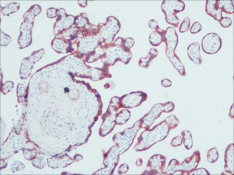
Anti-Cytokeratin 18 Antibody, clone RGE53 is an antibody against Cytokeratin 18 for use in FC, IC, IH & WB.
Research Category
Cell Structure
Research Sub Category
Cytokeratins
Western blot

Immunohistochemistry on fresh frozen or acetone fixed tissue sections. Formalin fixed, paraffin embedded tissues are not recommended for RGE53.

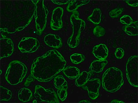
Immunocytochemistry: (10μg/mL) Light fixation with 4% formalin, 2.5% PFA ( 5-10 minutes room temperature) or acetone. {See, Barrett, et al 2001} Permeabilization with 0.2% triton X-100 in blocking buffer if using formalin or PFA fixations.

Flow cytometry: Acetone fixed/ethanol fixed cells {See Mezosi, E et al, 2002}.

Optimal working dilutions must be determined by the end user.

Target description

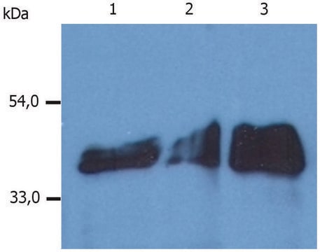
45 kD

Linkage

Replaces: 04-586

Physical form

Format: Purified
Protein A Purified mouse immunoglobulin in 20 mM sodium phosphate, 250 mM NaCl, pH. 7.6, with 0.1% sodium azide as a preservative.
Protein A purified

Storage and Stability

Maintain for 1 year at -20°C from date of shipment. Aliquot to avoid repeated freezing and thawing. For maximum recovery of product, centrifuge the original vial after thawing and prior to removing the cap.

Analysis Note

Control
Skin , Pancreas/tumor

Other Notes

Concentration: Please refer to the Certificate of Analysis for the lot-specific concentration.

Legal Information

CHEMICON is a registered trademark of Merck KGaA, Darmstadt, Germany

Disclaimer

Unless otherwise stated in our catalog or other company documentation accompanying the product(s), our products are intended for research use only and are not to be used for any other purpose, which includes but is not limited to, unauthorized commercial uses, in vitro diagnostic uses, ex vivo or in vivo therapeutic uses or any type of consumption or application to humans or animals.

Not finding the right product?  

Try our Product Selector Tool.

Storage Class Code

12 - Non Combustible Liquids

WGK

WGK 2

Flash Point(F)

Not applicable

Flash Point(C)

Not applicable


Certificates of Analysis (COA)

Search for Certificates of Analysis (COA) by entering the products Lot/Batch Number. Lot and Batch Numbers can be found on a product’s label following the words ‘Lot’ or ‘Batch’.

Already Own This Product?

Find documentation for the products that you have recently purchased in the Document Library.

Visit the Document Library

V Ninichuk et al.
Kidney international, 70(1), 121-129 (2006-05-26)
Multipotent mesenchymal stem or stromal cells (MSC) have shown to improve outcome of acute renal injury models, but whether MSC can delay renal failure in chronic kidney disease is not known. We injected primary MSC or saline into mice that
Sandra S Essbauer et al.
Virology journal, 8, 339-339 (2011-07-07)
Approximately 60% of emerging viruses are of zoonotic origin, with three-fourths derived from wild animals. Many of these zoonotic diseases are transmitted by rodents with important information about their reservoir dynamics and pathogenesis missing. One main reason for the gap
Noushin Amirpour et al.
Advanced biomedical research, 3, 4-4 (2014-03-05)
Retinal pigment epithelium (RPE) is a hexagonal monolayer of pigmented cells located between the neural retina and the choroid with an essential role for visual function. So, isolation, propagation and maintenance of their functional integrity of RPE are crucial for
Mustafa Şahiner et al.
Turkish journal of ophthalmology, 48(4), 190-195 (2018-09-12)
To assess the effects of anti-vascular endothelial growth factor (VEGF) drugs on retinal pigment epithelium cell culture. Aflibercept (0.5 mg/mL), bevacizumab (0.3125 mg/mL), and ranibizumab (0.125 mg/mL) were applied to retinal pigment epithelium cell cultures isolated from the enucleated eyes
K L Barrett et al.
The journal of histochemistry and cytochemistry : official journal of the Histochemistry Society, 49(7), 821-832 (2001-06-19)
In an earlier article from this laboratory, the current methods developed to detect apoptosis in cells and tissues were highlighted, along with the challenges in their interpretation. Recent discoveries concerning the underlying biochemical mechanisms of apoptotic effector pathways have made

Our team of scientists has experience in all areas of research including Life Science, Material Science, Chemical Synthesis, Chromatography, Analytical and many others.

Contact Technical Service