MAB131873
Anti-mCherry Antibody, clone 1C51
clone 1C51, from mouse
Sign Into View Organizational & Contract Pricing
All Photos(2)
About This Item
UNSPSC Code:
12352203
eCl@ss:
32160702
NACRES:
NA.41
Recommended Products
biological source
mouse
Quality Level
antibody form
purified antibody
antibody product type
primary antibodies
clone
1C51, monoclonal
species reactivity (predicted by homology)
all
technique(s)
immunocytochemistry: suitable
western blot: suitable
isotype
IgG2aκ
UniProt accession no.
shipped in
ambient
target post-translational modification
unmodified
General description
mCherry, red fluorescent protein, is derived from DsRed, a red fluorescent protein from disc corals of Discosoma species. It is monomeric form that has been engineered with multiple cycles of mutation, directed modification and evolutionary selection. It exhibits excitation maximum at 587 nm and an emission maximum at 610 nm with a fluorescent quantum yield of 0.22. mCherry displays high photostability and resistant to photobleaching. It also displays improved brightness and extremely rapid maturation rate. Anti-mCherry, clone1C51 is an affinity purified antibody that recognizes mCherry in HEK293 cells transfected with pFin-EF1-mCherry. (Ref.: Schaner, NC et al. (2004). Nat. Biotechnol. 22(12):1567-72).
Specificity
Clone 1C51 detects mCherry protein in multiple species.
Immunogen
His-tagged full length recombinant mCherry.
Application
Anti-mCherry Antibody, clone IC51, Cat. No. MAB131873 detects mCherry protein and is tested for use in Immunocytochemistry and Western Blotting.
Western Blotting Analysis: A representative lot detected mCherry in Western Blotting applications (Krajcovic, M., et. al. (2013). Mol Biol Cell. 24(23):3736-45).
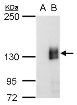
Western Blotting Analysis: A representative lot detected mCherry in HEK293 cells transfected with pFin-EF1-mCherry vs HEK293 untransfected cells (Courtesy of Dr Gerry Shaw).
Immunocytochemistry Analysis: A representative lot detected mCherry in HEK293 cells transfected with pFin-EF1-mCherry (Courtesy of Dr Gerry Shaw).
Quality
Evaluated by Western Blotting in HEK293 cells transfected with pFin-EF1-mCherry.
Western Blotting Analysis: 1 µg/mL of this antibody detected mCherry in 10 µg of lysate from HEK293 cells transfected with pFin-EF1-mCherry.
Western Blotting Analysis: 1 µg/mL of this antibody detected mCherry in 10 µg of lysate from HEK293 cells transfected with pFin-EF1-mCherry.
Target description
~32 kDa Observed; 26.72 kDa calculated. Uncharacterized bands may be observed in some lysate(s).
Physical form
Format: Purified
Purified mouse monoclonal antibody IgG2a in buffer containing PBS with 0.03% sodium azide with 50% glycerol.
Other Notes
Concentration: Please refer to lot specific datasheet.
Not finding the right product?
Try our Product Selector Tool.
Storage Class Code
10 - Combustible liquids
WGK
WGK 2
Certificates of Analysis (COA)
Search for Certificates of Analysis (COA) by entering the products Lot/Batch Number. Lot and Batch Numbers can be found on a product’s label following the words ‘Lot’ or ‘Batch’.
Already Own This Product?
Find documentation for the products that you have recently purchased in the Document Library.
Hongyan Sun et al.
iScience, 27(6), 110113-110113 (2024-07-02)
The gut epithelium is subject to constant renewal, a process reliant upon intestinal stem cell (ISC) proliferation that is driven by Wnt/β-catenin signaling. Despite the importance of Wnt signaling within ISCs, the relevance of Wnt signaling within other gut cell
N Quan et al.
Frontiers in genetics, 14, 1198203-1198203 (2023-09-25)
Protein misfolding is a common intracellular occurrence. Most mutations to coding sequences increase the propensity of the encoded protein to misfold. These misfolded molecules can have devastating effects on cells. Despite the importance of protein misfolding in human disease and
Brittany M White-Mathieu et al.
Current protocols, 4(5), e1051-e1051 (2024-05-23)
Fluorescent imaging of cellular membranes is challenged by the size of lipid bilayers, which are smaller than the diffraction limit of light. Recently, expansion microscopy (ExM) has emerged as an approachable super-resolution method that requires only widely accessible confocal microscopes.
Nicole L Nuckolls et al.
PLoS genetics, 18(12), e1009847-e1009847 (2022-12-09)
Meiotic drivers bias gametogenesis to ensure their transmission into more than half the offspring of a heterozygote. In Schizosaccharomyces pombe, wtf meiotic drivers destroy the meiotic products (spores) that do not inherit the driver from a heterozygote, thereby reducing fertility.
Memoona Ramzan et al.
Human mutation, 42(10), 1321-1335 (2021-07-16)
Hereditary deafness is clinically and genetically heterogeneous. We investigated deafness segregating as a recessive trait in two families. Audiological examinations revealed an asymmetric mild to profound hearing loss with childhood or adolescent onset. Exome sequencing of probands identified a homozygous
Our team of scientists has experience in all areas of research including Life Science, Material Science, Chemical Synthesis, Chromatography, Analytical and many others.
Contact Technical Service


