Skip to Content
Merck
All Photos(2)

Documents

L8271

Sigma-Aldrich

Monoclonal Anti-Laminin antibody produced in mouse

clone LAM-89, ascites fluid

Synonym(s):

Anti-Laminin Antibody

Sign Into View Organizational & Contract Pricing


About This Item

MDL number:
UNSPSC Code:
12352203
NACRES:
NA.41

biological source

mouse

Quality Level

conjugate

unconjugated

antibody form

ascites fluid

antibody product type

primary antibodies

clone

LAM-89, monoclonal

contains

15 mM sodium azide

species reactivity

feline, human, pig

should not react with

rabbit, lizard, sheep, carp, canine, chicken, rat, guinea pig, goat, frog, snake

technique(s)

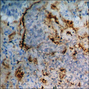
immunohistochemistry (formalin-fixed, paraffin-embedded sections): 1:1,000 using enzyme-treated human tissue sections
indirect ELISA: suitable
western blot: suitable

isotype

IgG1

UniProt accession no.

shipped in

dry ice

storage temp.

−20°C

target post-translational modification

unmodified

General description

Laminin, a 900kD (approx.) complex extracellular glycoprotein, is the most abundant structural and biologically active component present in the basement membranes. It is a cell substrate adhesion protein and thus has a crucial role in promoting neurite regeneration and modulating cellular functions like differentiation, cell shape and cell movement. Monoclonal Anti-laminin antibody can be used in immunohistochemistry to classify various disease processes involving basement membranes, to mark blood vessel walls of different species and to determine the origin and classification of human tumours. It can also be used in dot blot assays. Mouse anti- laminin antibody reacts specifically with purified human laminin but not with collagen IV, fibronectin, vitronectin or chondroitin sulfate types A, B, and C.
Monoclonal Anti-Laminin (mouse IgG1 isotype) is derived from the hybridoma produced by the fusion of mouse myeloma cells and splenocytes from an immunized mouse. Laminin, the most abundant structural and biologically active component in basement membranes, is a complex extracellular glycoprotein with an approximate molecular weight of 900 kDa. Laminin is composed of one A chain (400 kDa) one B1 chain (215 kDa) and one B2 chain (205 kDa) all held together by disulfide bonds. Laminin is only found in significant quantities in basement membranes, the thin extracellular matrices that surround epithelial tissue, nerve, fat cells and smooth, striated and cardiac muscle.

Immunogen

human laminin.

Application

Monoclonal Anti-Laminin antibody produced in mouse has been used in:
  • immunohistochemistry to stain matrix proteins
  • double immunofluorescence staining
  • indirect immunofluorescence
  • immunogold labelling for electron microscopy

Monoclonal Anti-laminin antibody can be used in indirect immunostaining to determine the distribution of laminin. It may also be used for ELISA, Immunofluorescence and western blotting.

Biochem/physiol Actions

Laminin is a cell substrate adhesion protein and thus has a crucial role in promoting neurite regeneration and modulating cellular functions like differentiation, cell shape and cell movement because it appears to be an important cell substrate adhesion protein. It plays an important role in many aspects of the cell biology. Variations in the expression of this protein have been observed in embryogenesis, organogenesis, post traumatic healing and cancer.

Disclaimer

Unless otherwise stated in our catalog or other company documentation accompanying the product(s), our products are intended for research use only and are not to be used for any other purpose, which includes but is not limited to, unauthorized commercial uses, in vitro diagnostic uses, ex vivo or in vivo therapeutic uses or any type of consumption or application to humans or animals.

Not finding the right product?  

Try our Product Selector Tool.

Storage Class Code

12 - Non Combustible Liquids

WGK

nwg

Flash Point(F)

Not applicable

Flash Point(C)

Not applicable


Certificates of Analysis (COA)

Search for Certificates of Analysis (COA) by entering the products Lot/Batch Number. Lot and Batch Numbers can be found on a product’s label following the words ‘Lot’ or ‘Batch’.

Already Own This Product?

Find documentation for the products that you have recently purchased in the Document Library.

Visit the Document Library

A Tezcaner et al.
Journal of biomedical materials research. Part A, 86(1), 170-181 (2007-10-25)
In developed countries the aging population faces increasing risks of blinding retinal diseases, for which there are few effective treatments available. Photoreceptor transplantation represents one approach, but generally results have been disappointing. We hypothesize that micropatterned biodegradable poly(L-lactic acid-co-glycolic acid)/poly(hydroxybutyrate-co-hydroxyvaleric
Andrea Farini et al.
Development (Cambridge, England), 143(4), 658-669 (2016-02-18)
Duchenne muscular dystrophy (DMD) is a progressive neuromuscular disorder characterized by muscle wasting and premature death. The defective gene is dystrophin, a structural protein, absence of which causes membrane fragility and myofiber necrosis. Several lines of evidence showed that in
Laminin-211 in skeletal muscle function
Holmberg J, et al.
Cell adhesion & migration, 7(1), 111-111 (2013)
Enhanced expression of matrilysin, collagenase, and stromelysin-1 in gastrointestinal ulcers.
Saarialho-Kere UK, et al.
The American Journal of Pathology, 148(2), 519-519 (1996)
Bryon R McKay et al.
The Journal of physiology, 588(Pt 17), 3307-3320 (2010-07-14)
In humans, muscle satellite cell (SC) enumeration is an important measurement used to determine the myogenic response to various stimuli. To date, the standard practice for enumeration is immunohistochemistry (IHC) using antibodies against common SC markers (Pax7, NCAM). Flow cytometry

Our team of scientists has experience in all areas of research including Life Science, Material Science, Chemical Synthesis, Chromatography, Analytical and many others.

Contact Technical Service