Skip to Content
Merck
All Photos(7)

Documents

HPA004832

Sigma-Aldrich

Anti-ARPC1B antibody produced in rabbit

enhanced validation

Prestige Antibodies® Powered by Atlas Antibodies, affinity isolated antibody, buffered aqueous glycerol solution

Synonym(s):

Anti-Actin-related protein 2/3 complex subunit 1B, Anti-Arp2/3 complex 41 kDa subunit, Anti-p41-ARC

Sign Into View Organizational & Contract Pricing


About This Item

UNSPSC Code:
12352203
Human Protein Atlas Number:
NACRES:
NA.43

biological source

rabbit

Quality Level

conjugate

unconjugated

antibody form

affinity isolated antibody

antibody product type

primary antibodies

clone

polyclonal

product line

Prestige Antibodies® Powered by Atlas Antibodies

form

buffered aqueous glycerol solution

species reactivity

human, mouse, rat

enhanced validation

orthogonal RNAseq
Learn more about Antibody Enhanced Validation

technique(s)

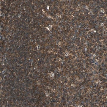
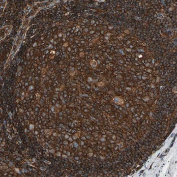
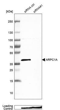
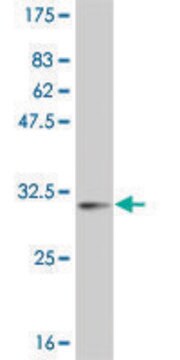
immunoblotting: 0.04-0.4 μg/mL
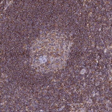
immunohistochemistry: 1:200-1:500

immunogen sequence

TVCLADADKKMAVATLASETLPLLALTFITDNSLVAAGHDCFPVLFTYDAAAGMLSFGGRLDVPKQSSQRGLTARERFQNLDKKASSEGGTAAGAGLDSLHKNSVSQISVLSGGKAKCSQFCTTGMDGGMSIWDVKSLESA

UniProt accession no.

shipped in

wet ice

storage temp.

−20°C

target post-translational modification

unmodified

Gene Information

human ... ARPC1B(10095)

General description

The Arp2/3 complex is composed of seven subunits, such as Arp2, Arp3, p41-Arc, p34-Arc, p21-Arc, p20-Arc, and p16-Arc (p complex). Among all the subunits, ARPC1B is a novel member of the Sop2 family of WD (tryptophan and aspartate) repeat-containing proteins.

Immunogen

Actin-related protein 2/3 complex subunit 1B recombinant protein epitope signature tag (PrEST)

Application

Applications in which this antibody has been used successfully, and the associated peer-reviewed papers, are given below.
Western Blotting (1 paper)

Biochem/physiol Actions

ARPC1B (actin related protein 2/3 complex, subunit 1B, 41kDa) is involved in the formation of actin filamentous network at the cell cortex. The protein subunit (ARPC1B) may maintain the structure and activity of the Arp2/3 complex. Overexpression of p41-Arc, a regulatory component of Arp2/3 complex causes the alteration of actin filaments. It gets accumulated in nuclei. Further, this alteration can trigger an intrinsic premature senescence in the tumor suppression process.

Features and Benefits

Prestige Antibodies® are highly characterized and extensively validated antibodies with the added benefit of all available characterization data for each target being accessible via the Human Protein Atlas portal linked just below the product name at the top of this page. The uniqueness and low cross-reactivity of the Prestige Antibodies® to other proteins are due to a thorough selection of antigen regions, affinity purification, and stringent selection. Prestige antigen controls are available for every corresponding Prestige Antibody and can be found in the linkage section.

Every Prestige Antibody is tested in the following ways:
  • IHC tissue array of 44 normal human tissues and 20 of the most common cancer type tissues.
  • Protein array of 364 human recombinant protein fragments.

Linkage

Corresponding Antigen APREST86831

Physical form

Solution in phosphate-buffered saline, pH 7.2, containing 40% glycerol and 0.02% sodium azide

Legal Information

Prestige Antibodies is a registered trademark of Merck KGaA, Darmstadt, Germany

Disclaimer

Unless otherwise stated in our catalog or other company documentation accompanying the product(s), our products are intended for research use only and are not to be used for any other purpose, which includes but is not limited to, unauthorized commercial uses, in vitro diagnostic uses, ex vivo or in vivo therapeutic uses or any type of consumption or application to humans or animals.

Not finding the right product?  

Try our Product Selector Tool.

Storage Class Code

10 - Combustible liquids

WGK

WGK 1

Flash Point(F)

Not applicable

Flash Point(C)

Not applicable

Personal Protective Equipment

dust mask type N95 (US), Eyeshields, Gloves

Certificates of Analysis (COA)

Search for Certificates of Analysis (COA) by entering the products Lot/Batch Number. Lot and Batch Numbers can be found on a product’s label following the words ‘Lot’ or ‘Batch’.

Already Own This Product?

Find documentation for the products that you have recently purchased in the Document Library.

Visit the Document Library

Yolla German et al.
Cell reports, 36(1), 109318-109318 (2021-07-08)
The immunological synapse is a complex structure that decodes stimulatory signals into adapted lymphocyte responses. It is a unique window to monitor lymphocyte activity because of development of systematic quantitative approaches. Here we demonstrate the applicability of high-content imaging to
Annabelle G Small et al.
Frontiers in immunology, 13, 840510-840510 (2022-03-24)
The phagocytosis-promoting complement receptor, Complement Receptor Immunoglobulin (CRIg), is exclusively expressed on macrophages. It has been demonstrated that expression in macrophages could be modulated by inflammatory mediators, including cytokines. This raised the possibility that a major phagocyte, the neutrophil, may
Yaser Gamallat et al.
International journal of molecular sciences, 23(3) (2022-02-16)
ARPC1B (Actin Related Protein 2/3 Complex Subunit 1B) has been found to be involved in platelet abnormalities of immune-mediated inflammatory disease and eosinophilia. However, its role in prostate cancer (PCa) has not been established. We characterized the role of ARPC1B
Un Jung Yun et al.
Experimental & molecular medicine, 43(7), 389-392 (2011-06-02)
Cellular senescence is a tumor-suppressive process instigated by proliferation in the absence of telomere replication, by cellular stresses such as oncogene activation, or by activation of the tumor suppressor proteins, such as Rb or p53. This process is characterized by
Fred G Pluthero et al.
Methods in molecular biology (Clifton, N.J.), 1812, 13-31 (2018-09-02)
Microscopy is central to studies of platelets and their precursor megakaryocytes. Here we describe methods to rapidly obtain high resolution images of fixed platelets, megakaryocytes and megakaryocytic cells via immunofluorescence microscopy. Protocols covered include: (1) isolation and preparation of cells

Our team of scientists has experience in all areas of research including Life Science, Material Science, Chemical Synthesis, Chromatography, Analytical and many others.

Contact Technical Service