Skip to Content
Merck
All Photos(1)

Key Documents

MABF3038

Sigma-Aldrich

Anti-Rhinovirus Capsid VP1 Antibody, clone HRV-18003

Synonym(s):

HRV-capsid VP1

Sign Into View Organizational & Contract Pricing


About This Item

UNSPSC Code:
12352203
NACRES:
NA.41
clone:
HRV-18003, monoclonal
application:
ELISA
WB
inhibition assay
species reactivity:
virus
technique(s):
ELISA: suitable
inhibition assay: suitable
western blot: suitable
citations:

biological source

mouse

Quality Level

antibody form

purified antibody

antibody product type

primary antibodies

clone

HRV-18003, monoclonal

mol wt

calculated mol wt 242.824 kDa
observed mol wt ~32 kDa

purified by

using protein G

species reactivity

virus

packaging

antibody small pack of 100

technique(s)

ELISA: suitable
inhibition assay: suitable
western blot: suitable

isotype

IgG2aκ

epitope sequence

N-terminal half

Protein ID accession no.

UniProt accession no.

storage temp.

-10 to -25°C

Specificity

Clone HRV-18003 is a mouse monoclonal antibody that detects Rhinovirus. It targets an epitope within 8 amino acids from the N-terminal half.

Immunogen

DNA encoding P1-2A and 3C of different strains of Rhinoviruses in pCAGGS plasmids.

Application

Quality Control Testing

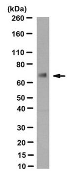
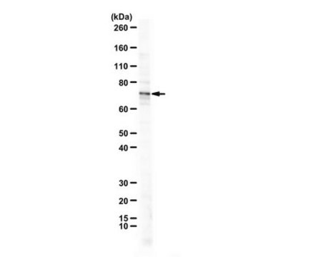
Evaluated by Western Blotting with recombinant Rhinovirus Capsid VP1 protein.

Western Blotting Analysis: A 1:125 dilution of this antibody detected recombinant Rhinovirus Capsid VP1 protein.

Tested Applications

Functional Assay A representative lot of this antibody exhibited strong activity in in vitro antibody dependent cellular phagocytosis (ADCP) assay. (Behzadi, M.A., et al. (2020). Sci Rep.;10(1):9750).

Western Blotting Analysis: A representative lot detected HRV-18003 recognizes VP1 in Western Blotting applications (Behzadi, M.A., et al. (2020). Sci Rep.;10(1):9750).

ELISA Analysis: A representative lot detected HRV-18003 recognizes VP1 in ELISA applications (Behzadi, M.A., et al. (2020). Sci Rep.;10(1):9750).

Note: Actual optimal working dilutions must be determined by end user as specimens, and experimental conditions may vary with the end user.

Target description

Rhinoviruses (RV) belong to the Picornavirus family and are known as a leading cause of respiratory infections. Over 160 different types of rhinoviruses have been identified and based on their genetic diversity and phylogenetic sequence analysis they are grouped as rhinovirus A, B, and C. Three different membrane glycoproteins are known to serve as receptors for these viruses. ICAM-1 is used by a majority of A group and all B group rhinoviruses, a small number of group A viruses use the low-density lipoprotein receptor (LDLR), and the group C of rhinoviruses use cadherin-related family member 3 receptor (CDKR3). Canyons on the surface of the virus are known to provide an attachment site for receptors on the surface of susceptible target cells. Viral infectivity is neutralized when host immunoglobulin G binds to the surface of the virus, thereby blocking interaction between the host cell receptor and the receptor binding site located at the base of the canyon. The RV is composed of single-stranded RNA within a capsid with icosahedral symmetry. The viral capsid of human RV is comprised of four viral proteins: VP1, VP2, VP3, and VP4. The remaining viral proteins are responsible for viral replication and subsequent assembly. Capsid protein VP1 forms an icosahedral capsid of pseudo-T=3 symmetry with capsid proteins VP2 and VP3. VP1 interacts with host cell receptor to provide virion attachment to target host cells. This attachment induces virion internalization. VP1 is the most exposed surface protein and plays a critical role in viral antigenicity and induction of neutralizing antibodies. Clone HRV-18003 binds to a conserve epitope within VP1 and displays cross reactivity with multiple types of rhinoviruses, including A1A, A1B, A15, and A49. It exhibits strong antibody dependent cellular phagocytosis (ADCP) against A1A and A16 viruses. (Ref.: Behzadi, MA., et al. (2020). Sci. Rep. 10(1):9750; Basnet, S., et al. (2019). Chest. ;155(5):1018-1025).

Physical form

Purified mouse monoclonal antibody IgG2a in PBS without preservatives.

Reconstitution

0.5 mg/mL. Please refer to guidance on suggested starting dilutions and/or titers per application and sample type.

Storage and Stability

Store at -10°C to -25°C. Handling Recommendations: Upon receipt and prior to removing the cap, centrifuge the vial and gently mix the solution. Aliquot into microcentrifuge tubes and store at -20°C. Avoid repeated freeze/thaw cycles, which may damage IgG and affect product performance.

Other Notes

Concentration: Please refer to the Certificate of Analysis for the lot-specific concentration.

Disclaimer

Unless otherwise stated in our catalog or other company documentation accompanying the product(s), our products are intended for research use only and are not to be used for any other purpose, which includes but is not limited to, unauthorized commercial uses, in vitro diagnostic uses, ex vivo or in vivo therapeutic uses or any type of consumption or application to humans or animals.

Not finding the right product?  

Try our Product Selector Tool.

Storage Class Code

12 - Non Combustible Liquids

WGK

WGK 2

Flash Point(F)

Not applicable

Flash Point(C)

Not applicable


Certificates of Analysis (COA)

Search for Certificates of Analysis (COA) by entering the products Lot/Batch Number. Lot and Batch Numbers can be found on a product’s label following the words ‘Lot’ or ‘Batch’.

Already Own This Product?

Find documentation for the products that you have recently purchased in the Document Library.

Visit the Document Library

Our team of scientists has experience in all areas of research including Life Science, Material Science, Chemical Synthesis, Chromatography, Analytical and many others.

Contact Technical Service