Skip to Content
Merck
All Photos(1)

Documents

ABE285

Sigma-Aldrich

Anti-Tristetraprolin Antibody

from rabbit, purified by affinity chromatography

Synonym(s):

Tristetraprolin, TTP, G0/G1 switch regulatory protein 24, Growth factor-inducible nuclear protein NUP475, Protein TIS11A, TIS11, Zinc finger protein 36 homolog, Zfp-36

Sign Into View Organizational & Contract Pricing


About This Item

UNSPSC Code:
12352203
eCl@ss:
32160702
NACRES:
NA.41

biological source

rabbit

Quality Level

antibody form

affinity isolated antibody

antibody product type

primary antibodies

clone

polyclonal

purified by

affinity chromatography

species reactivity

human, mouse

technique(s)

RIP: suitable
immunocytochemistry: suitable
immunoprecipitation (IP): suitable
western blot: suitable

NCBI accession no.

UniProt accession no.

shipped in

wet ice

target post-translational modification

unmodified

Gene Information

human ... ZFP36(7538)

General description

Tristetraprolin (TTP, Nup 475, TIS11, G0S24, or ZFP36) is a member of the CCCH tandem zinc finger protein family. It is a nuclear RNA-binding protein that functions as a trans-acting factor. It is involved in post-transcriptional regulation of cytokine genes, most notably Tumor Necrosis Factor alpha (TNF-α). Upon activation by lipopolysaccharides, TTP binds TNF-α mRNA at AU-rich domains in the 3′-untranslated Region (3′-UTR) and induces deadenylation, thereby destabilizing the mRNA and tagging it for degradation. To produce this effect, TTP recruits other trans-acting factors to its C-terminal domain and mRNA-decay enzymes to its N-terminal domain. TTP is deactivated by phosphorylation driven by mitogen-activated protein kinase-activated protein kinase 2 (MK2) and p53. Therefore, TTP/MK2 regulates the biosynthesis of TNF-α and mediates inflammatory responses involving TNF-α. Knockout of TTP in mice produces arthritis, alopecia, dermatitis, among a range of other phenotypes related to overexpression of TNF-α.

Immunogen

KLH-conjugated linear peptide corresponding to human Tristetraprolin.

Application

Anti-Tristetraprolin Antibody is an antibody against Tristetraprolin for use in Western Blotting, ICC, Immunoprecipitation, RNA Binding Protein Immunoprecipitation (RIP).
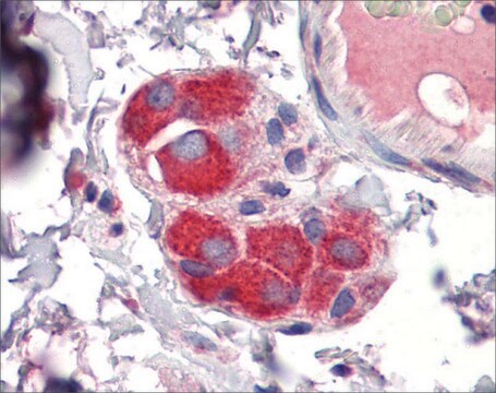
Immunocytochemistry Analysis: A representative lot was used by an independent laboratory to detect Tristetraprolin in human leukocytes. (Brooks, S., et al. (2002). Arthritis and Rheumatism. 46(5):1362–1370.)
Research Category
Epigenetics & Nuclear Function
Research Sub Category
Growth Factors & Receptors

Quality

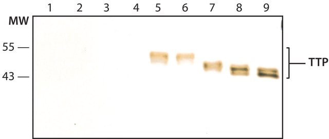
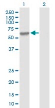
Evaluated by Western Blot in A431 cell lysate.

Western Blot Analysis: 1 µg/mL of this antibody detected Tristetraprolin on 10 µg of A431 cell lysate.

Target description

~40 kDa observed. Uniprot gives a calculated molecular weight of 34 kDa An uncharacterized band appears at ~53 kDa in some lysates.

Physical form

Affinity purified
Purified rabbit polyclonal in buffer containing PBS without 0.05% sodium azide.

Storage and Stability

Stable for 1 year at 2-8°C from date of receipt.

Analysis Note

Control
A431 cell lysate

Other Notes

Concentration: Please refer to the Certificate of Analysis for the lot-specific concentration.

Disclaimer

Unless otherwise stated in our catalog or other company documentation accompanying the product(s), our products are intended for research use only and are not to be used for any other purpose, which includes but is not limited to, unauthorized commercial uses, in vitro diagnostic uses, ex vivo or in vivo therapeutic uses or any type of consumption or application to humans or animals.

Not finding the right product?  

Try our Product Selector Tool.

Storage Class Code

12 - Non Combustible Liquids

WGK

WGK 2

Flash Point(F)

Not applicable

Flash Point(C)

Not applicable


Certificates of Analysis (COA)

Search for Certificates of Analysis (COA) by entering the products Lot/Batch Number. Lot and Batch Numbers can be found on a product’s label following the words ‘Lot’ or ‘Batch’.

Already Own This Product?

Find documentation for the products that you have recently purchased in the Document Library.

Visit the Document Library

Yuki Hikichi et al.
The Journal of experimental medicine, 218(12) (2021-10-29)
Group 2 innate lymphoid cells (ILC2s) are unique in their ability to produce low levels of type 2 cytokines at steady state, and their production capacity is dramatically increased upon stimulation with IL-33. However, it is unknown how constitutive cytokine
Jan Meier et al.
RNA biology, 10(6), 1018-1029 (2013-05-16)
MicroRNAs (miRNAs) are single-stranded, small, non-coding RNAs, which fine-tune protein expression by degrading and/or translationally inhibiting mRNAs. Manipulation of miRNA expression in animal models frequently results in severe phenotypes indicating their relevance in controlling cellular functions, most likely by interacting
Moritz Haneklaus et al.
The Journal of biological chemistry, 292(17), 6869-6881 (2017-03-18)
The NLRP3 inflammasome is a central regulator of inflammation in many common diseases, including atherosclerosis and type 2 diabetes, driving the production of pro-inflammatory mediators such as IL-1β and IL-18. Due to its function as an inflammatory gatekeeper, expression and
Yueh-Chien Lin et al.
Biomedicines, 10(3) (2022-03-26)
Prostate cancer (PCa) is the major cause of cancer-related death among aging men worldwide. Recent studies have suggested that calreticulin (CRT), a multifunctional chaperon protein, may play an important role in the regulation of PCa tumorigenesis and progression. However, the
Comparative phosphoproteomics reveals components of host cell invasion and post-transcriptional regulation during Francisella infection.
Nakayasu, ES; Tempel, R; Cambronne, XA; Petyuk, VA; Jones, MB; Gritsenko, MA; Monroe et al.
Molecular and Cellular Proteomics null

Our team of scientists has experience in all areas of research including Life Science, Material Science, Chemical Synthesis, Chromatography, Analytical and many others.

Contact Technical Service