Skip to Content
Merck
All Photos(2)

Documents

C2542

Sigma-Aldrich

Monoclonal Anti-N-Cadherin antibody produced in mouse

clone GC-4, ascites fluid

Synonym(s):

Anti-A-CAM, Anti-A-Cell Adhesion Molecule

Sign Into View Organizational & Contract Pricing


About This Item

MDL number:
UNSPSC Code:
12352203
NACRES:
NA.41

biological source

mouse

Quality Level

conjugate

unconjugated

antibody form

ascites fluid

antibody product type

primary antibodies

clone

GC-4, monoclonal

contains

15 mM sodium azide

species reactivity

chicken, mouse, human, rabbit, rat

technique(s)

electron microscopy: suitable
immunohistochemistry (frozen sections): 1:100 using chicken cardiac muscle
microarray: suitable
western blot: 1:100 using chicken or rat cardiac muscle

UniProt accession no.

application(s)

research pathology

shipped in

dry ice

storage temp.

−20°C

target post-translational modification

unmodified

Gene Information

human ... CDH2(1000)
mouse ... Cdh2(12558)
rat ... Cdh2(83501)

Looking for similar products? Visit Product Comparison Guide

General description

N-cadherin is a calcium dependent cell adhesion molecule and is present mainly in cancer cells. Monoclonal anti-N-cadherin antibody reacts with the N-terminal portion of the extracellular domain of adipocyte adhesion molecule (ACAM).

Specificity

Monoclonal anti-N-cadherin antibody reacts specifically with A-CAM molecule in human, mouse, rabbit, chicken and rat.

Immunogen

Affinity purified chicken heart A-CAM.

Application

Monoclonal anti-N-cadherin antibody can be used in immunofluorescent microscopy, electron microscopy and immunoblotting to study the receptor localization of intercellular adherens. It may also be used to study the mechanism of adhesion and interaction in tissues.
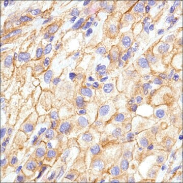
Monoclonal anti-N-cadherin antibody can be used in immunohistochemistry (1: 100) and as primary antibody in western blotting (1:1000) .

Biochem/physiol Actions

N-cadherin regulates the Src kinase pathway and also promotes cell motility and invasion. It inhibits adherens junction formation and disrupts existing junctions of cultured lens cells.

Physical form

Monoclonal Anti-N-Cadherin antibody produced in mouse is supplied as ascites fluid containing 15mM sodium azide.

Storage and Stability

For continuous use, store at 2-8 °C for up to one month. For extended storage, solution may be frozen in working aliquots. Repeated freezing and thawing is not recommended. If slight turbidity occurs upon prolonged storage, clarify by centrifugation before use.

Disclaimer

Unless otherwise stated in our catalog or other company documentation accompanying the product(s), our products are intended for research use only and are not to be used for any other purpose, which includes but is not limited to, unauthorized commercial uses, in vitro diagnostic uses, ex vivo or in vivo therapeutic uses or any type of consumption or application to humans or animals.

Not finding the right product?  

Try our Product Selector Tool.

Storage Class Code

10 - Combustible liquids

WGK

nwg

Flash Point(F)

Not applicable

Flash Point(C)

Not applicable


Certificates of Analysis (COA)

Search for Certificates of Analysis (COA) by entering the products Lot/Batch Number. Lot and Batch Numbers can be found on a product’s label following the words ‘Lot’ or ‘Batch’.

Already Own This Product?

Find documentation for the products that you have recently purchased in the Document Library.

Visit the Document Library

George Britton et al.
Methods in molecular biology (Clifton, N.J.), 2258, 119-130 (2020-12-20)
In the developing mammalian embryo, intercellular signaling allows cells to self-organize to create spatial patterns of different cell fates. This process is challenging to study because of the difficulty of observing or manipulating embryos on the spatial and temporal scales
C I Marín-Briggiler et al.
International journal of andrology, 33(1), e228-e239 (2009-10-21)
Neural cadherin (N-cadherin) is a transmembrane glycoprotein involved in calcium-dependent cell-cell adhesion and signalling events. The previous evidence shows N-cadherin expression in the human gonads and gametes; however, N-cadherin subcellular localization in human spermatozoa and oocytes, and its involvement in
Kamiel A J Kuijpers et al.
BioMed research international, 2014, 464217-464217 (2014-04-11)
Interstitial cells, also called myofibroblasts, most probably play a major role in the pathogenesis of the overactive bladder. However, no specific phenotypic marker has been identified. We investigated whether N-cadherin could play a role as a discriminatory marker for interstitial
Exogenous expression of N-cadherin in breast cancer cells induces cell migration, invasion, and metastasis
Hazan RB
The Journal of cell biology, 148(4), 779-790 (2000)
Kamiel A J Kuijpers et al.
BioMed research international, 2014, 754596-754596 (2014-05-16)
The human bladder shows spontaneous autonomous activity. Detrusor overactivity could be seen as a consequence of exaggerated autonomous activity. Interstitial cells (ICs) play a potential role in coordination of autonomous activity. As it is suggested that changes in ICs coexist

Our team of scientists has experience in all areas of research including Life Science, Material Science, Chemical Synthesis, Chromatography, Analytical and many others.

Contact Technical Service